 Explore
 Validate
 Learn
Learn Western blot
Antibody data
- Antibody Data
- Antigen structure
- References [0]
- Comments [0]
- Validations
- Western blot [4]
- Immunocytochemistry [3]
- Immunohistochemistry [1]
Submit
Validation data
Reference
Comment
Report error
- Product number
- PA5-20409 - Provider product page

- Provider
- Invitrogen Antibodies
- Product name
- TDP-43 Polyclonal Antibody
- Antibody type
- Polyclonal
- Antigen
- Synthetic peptide
- Description
- A suggested positive control is L1210 cell lysate. PA5-20409 can be used with blocking peptide PEP-0526.
- Reactivity
- Human, Mouse, Rat
- Host
- Rabbit
- Isotype
- IgG
- Vial size
- 100 µg
- Concentration
- 1 mg/mL
- Storage
- Maintain refrigerated at 2-8°C for up to 3 months. For long term storage store at -20°C
No comments: Submit comment
Supportive validation
- Submitted by
- Invitrogen Antibodies (provider)
- Main image
- Experimental details
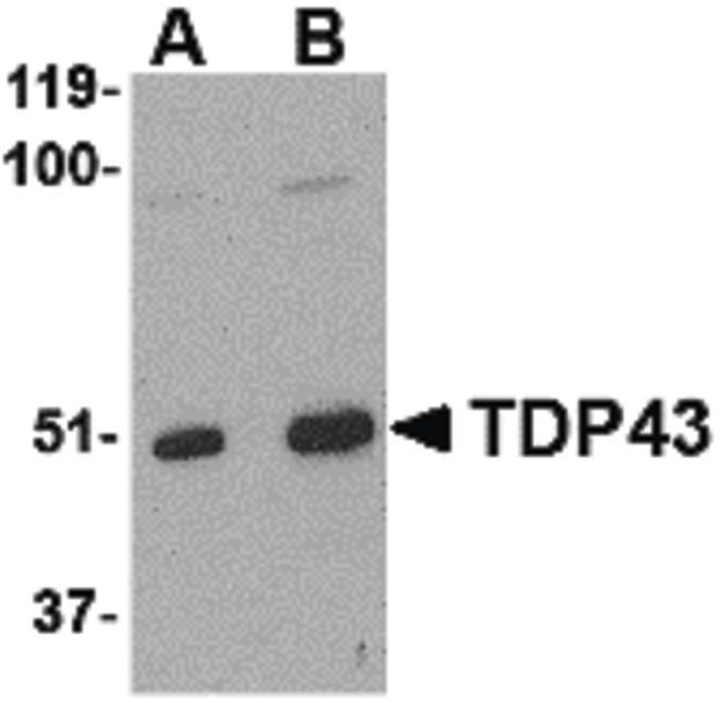
- Western blot analysis of L1210 cell lysate using a TDP43 polyclonal antibody (Product # PA5-20409) at (A) 0.5, (B) 1 and (C) 2 µg/mL.
- Submitted by
- Invitrogen Antibodies (provider)
- Main image
- Experimental details
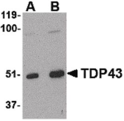
- Western Blot analysis of TDP43 in L1210 cell lysate with TDP-43 Polyclonal Antibody (Product # PA5-20409) at (A) 0.5 and (B) 1 µg/mL.
- Submitted by
- Invitrogen Antibodies (provider)
- Main image
- Experimental details
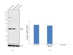
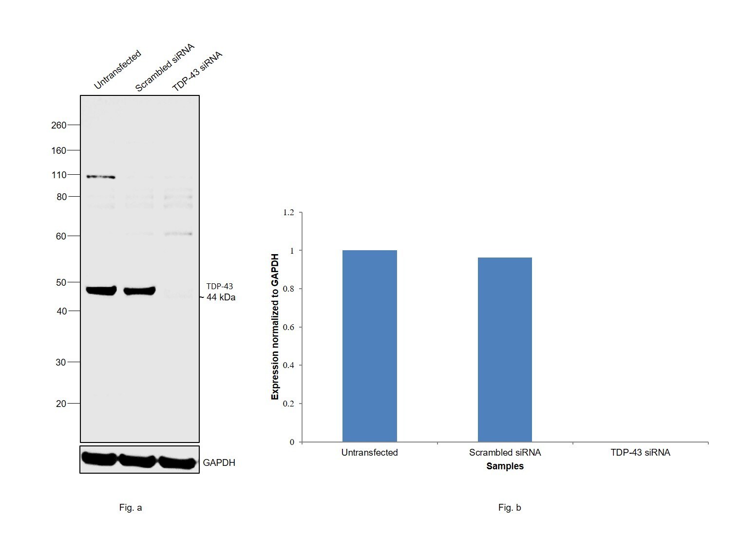
- Knockdown of TDP-43 was achieved by transfecting HeLa with TDP-43 specific siRNAs (Silencer® select Product # s23830, s23829). Western blot analysis (Fig. a) was performed using Whole Cell Extract-WCL from the TDP-43 knockdown cells (lane 3), non-targeting scrambled siRNA transfected cells (lane 2) and untransfected cells (lane 1). The blot was probed with TDP-43 Polyclonal Antibody (Product # PA5-20409, 0.5 ug/ml) and Goat anti-Rabbit IgG (H+L) Superclonal™ Recombinant Secondary Antibody, HRP (Product # A27036, 1:4000 dilution). Densitometric analysis of this western blot is shown in histogram (Fig. b). Decrease in signal upon siRNA mediated knock down confirms that antibody is specific to TDP-43.
- Submitted by
- Invitrogen Antibodies (provider)
- Main image
- Experimental details
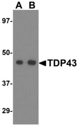
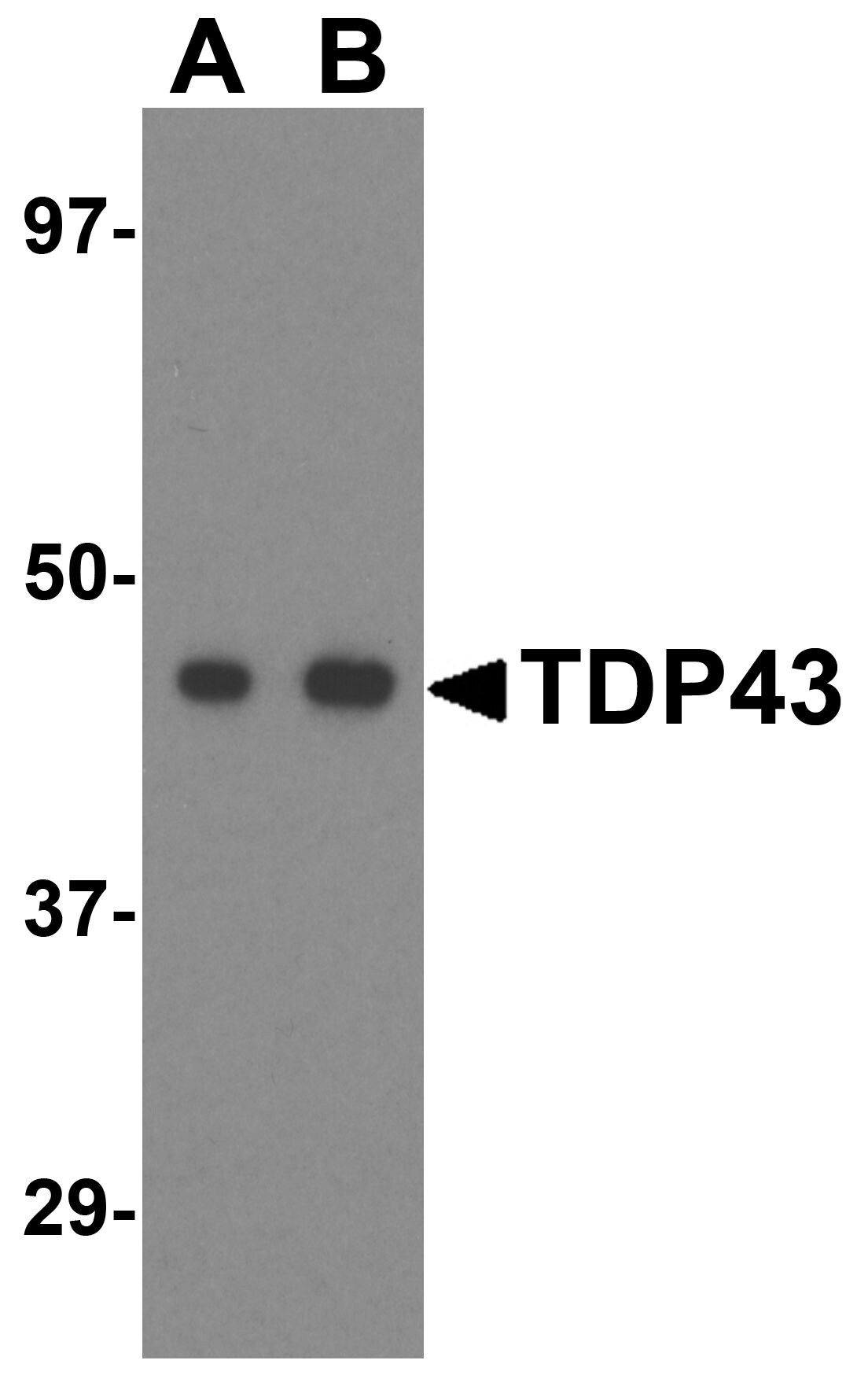
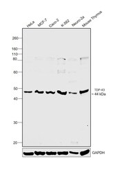
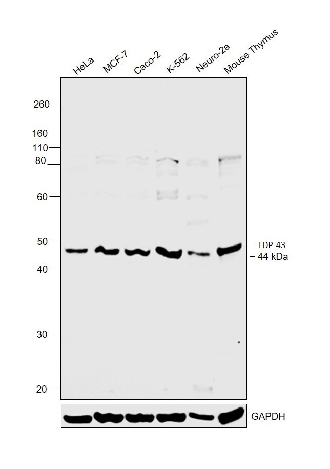
- Western blot was performed using Anti-TDP-43 Polyclonal Antibody(Product # PA5-20409) and a 44kDa band corresponding to TDP-43 was observed across cell lines and tissue extract tested. Whole Cell Extract-WCL (30 µg lysate) of HeLa (Lane 1), MCF7 (Lane 2), Caco-2 (Lane 3), K-562 (Lane 4), Neuro-2a (Lane 5) and tissue extract of Mouse Thymus (Lane 6) were electrophoresed using NuPAGE™ 4-12% Bis-Tris Protein Gel (Product # NP0322BOX). Resolved proteins were then transferred onto a Nitrocellulose membrane (Product # IB23001) by iBlot® 2 Dry Blotting System (Product # IB21001). The blot was probed with the primary antibody (0.5 µg/mL) and detected by chemiluminescence with Goat anti-Rabbit IgG (H+L) Superclonal™ Recombinant Secondary Antibody, HRP (Product # A27036,1:4000 dilution) using the iBright FL 1000 (Product # A32752). Chemiluminescent detection was performed using Novex® ECL Chemiluminescent Substrate Reagent Kit (Product # WP20005).
Supportive validation
- Submitted by
- Invitrogen Antibodies (provider)
- Main image

- Experimental details
- Immunofluorescent analysis of HeLa cells using a TDP43 polyclonal antibody (Product # PA5-20409) at a 20 µg/mL dilution.
- Submitted by
- Invitrogen Antibodies (provider)
- Main image

- Experimental details
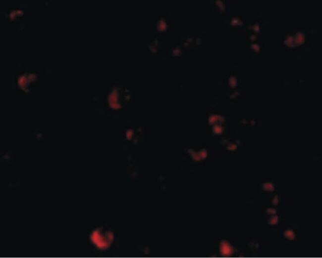
- Immunocytochemistry of TDP43 in HeLa cells with TDP-43 Polyclonal Antibody (Product # PA5-20409) at 5 µg/mL.
- Submitted by
- Invitrogen Antibodies (provider)
- Main image

- Experimental details
- Immunofluorescence of TDP43 in Hela cells with TDP-43 Polyclonal Antibody (Product # PA5-20409) at 20 µg/mL.

Supportive validation
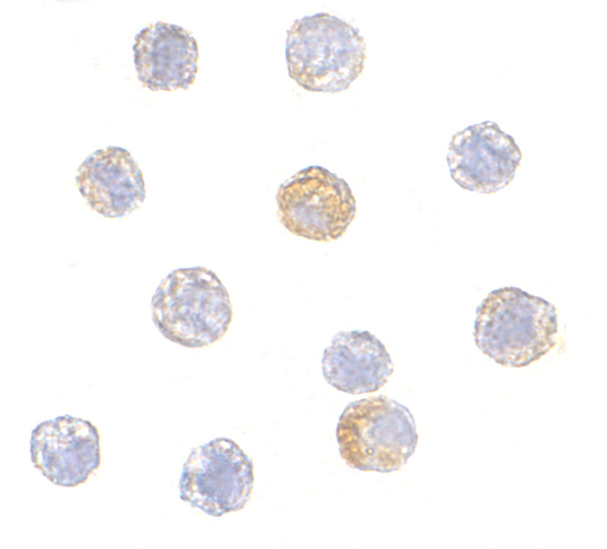
- Submitted by
- Invitrogen Antibodies (provider)
- Main image

- Experimental details
- Immunocytochemistry staining of HeLa cells using a TDP43 polyclonal antibody (Product # PA5-20409) at a 5 µg/mL dilution.
