Explore
 Validate
 Learn
LearnPA5-29949
antibody from Invitrogen Antibodies
Targeting: TARDBP
ALS10, TDP-43
 Western blot
 Immunocytochemistry Immunoprecipitation Immunohistochemistry
 Chromatin Immunoprecipitation Other assay
Antibody data
- Antibody Data
- Antigen structure
- References [3]
- Comments [0]
- Validations
- Immunocytochemistry [7]
- Immunoprecipitation [1]
- Immunohistochemistry [3]
- Other assay [3]
Submit
Validation data
Reference
Comment
Report error
- Product number
- PA5-29949 - Provider product page

- Provider
- Invitrogen Antibodies
- Product name
- TDP-43 Polyclonal Antibody
- Antibody type
- Polyclonal
- Antigen
- Recombinant full-length protein
- Description
- Recommended positive controls: 293T, A431, HeLa, HepG2, Neuro2A, GL261, NIH-3T3, BCL-1, Raw264.7, PC-12, Rat-2, Rat brain. Predicted reactivity: Mouse (95%), Rat (95%), Zebrafish (83%), Xenopus laevis (91%), Chicken (95%), Bovine (96%). Store product as a concentrated solution. Centrifuge briefly prior to opening the vial.
- Reactivity
- Human, Mouse, Rat
- Host
- Rabbit
- Isotype
- IgG
- Vial size
- 100 μL
- Concentration
- 0.63 mg/mL
- Storage
- Store at 4°C short term. For long term storage, store at -20°C, avoiding freeze/thaw cycles.
Submitted references The RNA m6A reader YTHDF2 controls NK cell antitumor and antiviral immunity.
Association of Position Played and Career Duration and Chronic Traumatic Encephalopathy at Autopsy in Elite Football and Hockey Players.
DNA repair deficiency and senescence in concussed professional athletes involved in contact sports.
Ma S, Yan J, Barr T, Zhang J, Chen Z, Wang LS, Sun JC, Chen J, Caligiuri MA, Yu J
The Journal of experimental medicine 2021 Aug 2;218(8)
The Journal of experimental medicine 2021 Aug 2;218(8)
Association of Position Played and Career Duration and Chronic Traumatic Encephalopathy at Autopsy in Elite Football and Hockey Players.
Schwab N, Wennberg R, Grenier K, Tartaglia C, Tator C, Hazrati LN
Neurology 2021 Apr 6;96(14):e1835-e1843
Neurology 2021 Apr 6;96(14):e1835-e1843
DNA repair deficiency and senescence in concussed professional athletes involved in contact sports.
Schwab N, Grenier K, Hazrati LN
Acta neuropathologica communications 2019 Nov 14;7(1):182
Acta neuropathologica communications 2019 Nov 14;7(1):182
No comments: Submit comment
Supportive validation
- Submitted by
- Invitrogen Antibodies (provider)
- Main image

- Experimental details
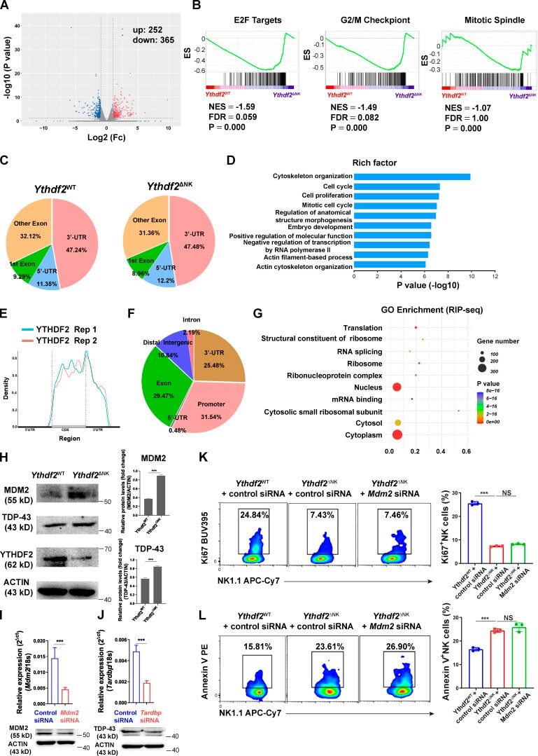
- Immunofluorescent analysis of TARDBP in paraformaldehyde-fixed HeLa cells using a TARDBP polyclonal antibody (Product # PA5-29949) at a 1:200 dilution.
- Submitted by
- Invitrogen Antibodies (provider)
- Main image
- Experimental details
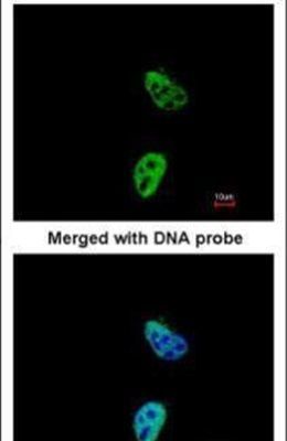
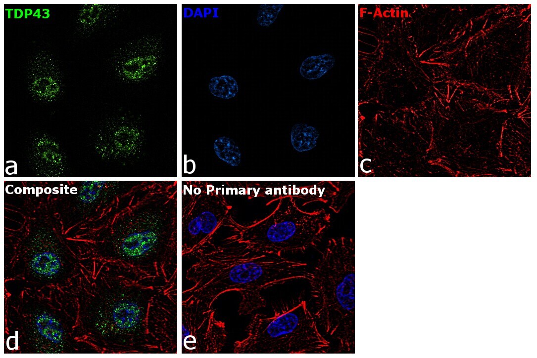
- TDP-43 Polyclonal Antibody detects TDP43 protein by immunofluorescent analysis. Sample: DIV10 rat E18 primary cortical neuron cells were fixed in 4% paraformaldehyde at RT for 15 min. Green: TDP43 stained by TDP-43 Polyclonal Antibody (Product # PA5-29949) diluted at 1:500. Red: Tau, stained by Phospho-Tau (Ser262) Polyclonal Antibody [GT287]diluted at 1:500. Blue: Fluoroshield with DAPI .

- Submitted by
- Invitrogen Antibodies (provider)
- Main image
- Experimental details
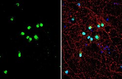
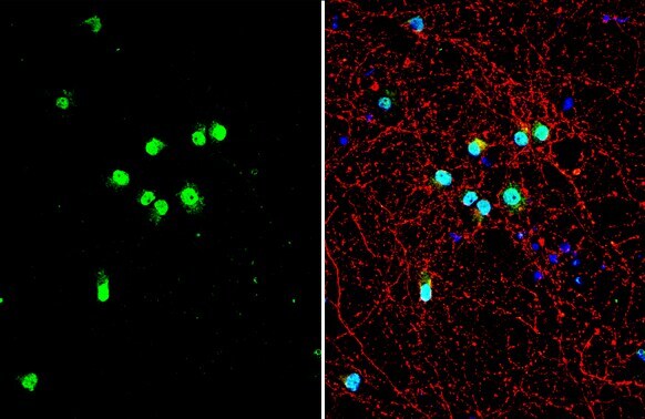
- TDP-43 Polyclonal Antibody detects TDP43 protein at nucleus by immunofluorescent analysis. Sample: HeLa cells were fixed in 4% paraformaldehyde at RT for 15 min. Green: TDP43 stained by TDP-43 Polyclonal Antibody (Product # PA5-29949) diluted at 1:2,000. Red: phalloidin, a cytoskeleton marker, diluted at 1:200.

- Submitted by
- Invitrogen Antibodies (provider)
- Main image
- Experimental details
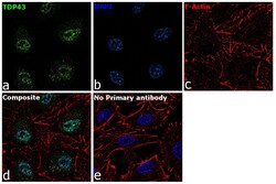
- Immunofluorescence analysis of TDP-43 was performed using 70% confluent log phase HeLa cells. The cells were fixed with 4% paraformaldehyde for 10 minutes, permeabilized with 0.1% Triton™ X-100 for 15 minutes, and blocked with 2% BSA for 45 minutes at room temperature. The cells were labeled with TDP-43 Polyclonal Antibody (Product # PA5-29949) at 5 µg/mL in 0.1% BSA, incubated at 4 degree celsius overnight and then labeled with Goat anti-Rabbit IgG (H+L) Highly Cross-Adsorbed Secondary Antibody, Alexa Fluor Plus 488 (Product # A32731), (1:2000), for 45 minutes at room temperature (Panel a: Green). Nuclei (Panel b:Blue) were stained with ProLong™ Diamond Antifade Mountant with DAPI (Product # P36962). F-actin (Panel c: Red) was stained with Rhodamine Phalloidin (Product # R415, 1:300). Panel d represents the merged image showing Nuclear localization. Panel e represents control cells with no primary antibody to assess background. The images were captured at 60X magnification.
- Submitted by
- Invitrogen Antibodies (provider)
- Main image

- Experimental details
- Immunofluorescence analysis of TDP-43 was performed using 70% confluent log phase HeLa cells. The cells were fixed with 4% paraformaldehyde for 10 minutes, permeabilized with 0.1% Triton™ X-100 for 15 minutes, and blocked with 2% BSA for 45 minutes at room temperature. The cells were labeled with TDP-43 Polyclonal Antibody (Product # PA5-29949) at 5 µg/mL in 0.1% BSA, incubated at 4 degree celsius overnight and then labeled with Goat anti-Rabbit IgG (H+L) Highly Cross-Adsorbed Secondary Antibody, Alexa Fluor Plus 488 (Product # A32731), (1:2000), for 45 minutes at room temperature (Panel a: Green). Nuclei (Panel b:Blue) were stained with ProLong™ Diamond Antifade Mountant with DAPI (Product # P36962). F-actin (Panel c: Red) was stained with Rhodamine Phalloidin (Product # R415, 1:300). Panel d represents the merged image showing Nuclear localization. Panel e represents control cells with no primary antibody to assess background. The images were captured at 60X magnification.

- Submitted by
- Invitrogen Antibodies (provider)
- Main image

- Experimental details
- TDP-43 Polyclonal Antibody detects TDP43 protein by immunofluorescent analysis. Sample: DIV10 rat E18 primary cortical neuron cells were fixed in 4% paraformaldehyde at RT for 15 min. Green: TDP43 stained by TDP-43 Polyclonal Antibody (Product # PA5-29949) diluted at 1:500. Red: Tau, stained by Phospho-Tau (Ser262) Polyclonal Antibody [GT287]diluted at 1:500. Blue: Fluoroshield with DAPI .
- Submitted by
- Invitrogen Antibodies (provider)
- Main image

- Experimental details
- TDP-43 Polyclonal Antibody detects TDP43 protein at nucleus by immunofluorescent analysis. Sample: HeLa cells were fixed in 4% paraformaldehyde at RT for 15 min. Green: TDP43 stained by TDP-43 Polyclonal Antibody (Product # PA5-29949) diluted at 1:2,000. Red: phalloidin, a cytoskeleton marker, diluted at 1:200.
Supportive validation
- Submitted by
- Invitrogen Antibodies (provider)
- Main image
- Experimental details
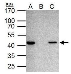
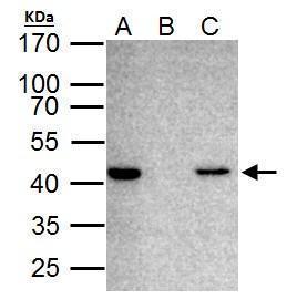
- TARDBP antibody immunoprecipitates TARDBP protein in IP experiments. IP Sample: HeLa whole cell lysate/extract A. 40 µg HeLa whole cell lysate/extract B. Control with 2 µg of preimmune rabbit IgG C. Immunoprecipitation of TARDBP protein by 2 µg of TARDBP antibody (Product # PA5-29949) 12% SDS-PAGE The immunoprecipitated TARDBP protein was detected by TARDBP antibody (Product # PA5-29949) diluted at 1:1,000.

Supportive validation
- Submitted by
- Invitrogen Antibodies (provider)
- Main image
- Experimental details
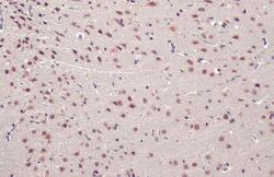
- TDP-43 Polyclonal Antibody detects TDP43 protein at nucleus by immunohistochemical analysis. Sample: Paraffin-embedded rat brain. TDP43 stained by TDP-43 Polyclonal Antibody (Product # PA5-29949) diluted at 1:500. Antigen Retrieval: Citrate buffer, pH 6.0, 15 min.
- Submitted by
- Invitrogen Antibodies (provider)
- Main image
- Experimental details
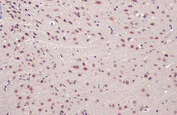
- TDP-43 Polyclonal Antibody detects TDP43 protein at nucleus by immunohistochemical analysis. Sample: Paraffin-embedded mouse brain. TDP43 stained by TDP-43 Polyclonal Antibody (Product # PA5-29949) diluted at 1:500. Antigen Retrieval: Citrate buffer, pH 6.0, 15 min.

- Submitted by
- Invitrogen Antibodies (provider)
- Main image
- Experimental details
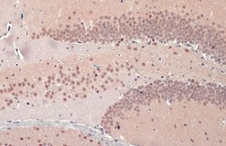
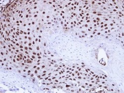
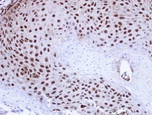
- Immunohistochemical analysis of paraffin-embedded Cal27 Xenograft , using TDP-43 (Product # PA5-29949) antibody at 1:100 dilution. Antigen Retrieval: EDTA based buffer, pH 8.0, 15 min.
Supportive validation
- Submitted by
- Invitrogen Antibodies (provider)
- Main image

- Experimental details
- TARDBP antibody immunoprecipitates TARDBP protein in IP experiments. IP Sample: HeLa whole cell lysate/extract A. 40 µg HeLa whole cell lysate/extract B. Control with 2 µg of preimmune rabbit IgG C. Immunoprecipitation of TARDBP protein by 2 µg of TARDBP antibody (Product # PA5-29949) 12% SDS-PAGE The immunoprecipitated TARDBP protein was detected by TARDBP antibody (Product # PA5-29949) diluted at 1:1,000.
- Submitted by
- Invitrogen Antibodies (provider)
- Main image

- Experimental details
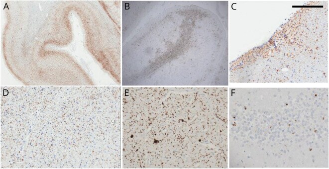
- Figure 2 Neuropathologic Changes in More Advanced Cases of Chronic Traumatic Encephalopathy Widespread cortical tau deposits in superficial cortical layers and involving both gyri and sulci in a continuous pattern. Some of the superficial cortical pathology can be focal and involve the crown of the gyri with widespread white matter punctate tau deposits in the (B and D) white matter and (C) subventricular areas. Other concomitant pathologies are alpha-synuclein deposits in the shape of Lewy bodies and neurites as depicted in the (E) cingulate cortex and (F) TAR DNA-binding protein 43 deposits as shown in the dentate gyrus. Scale bar in the top left corner represents 0.1 cm in panels A and B and 0.01 cm in panels C-F.
- Submitted by
- Invitrogen Antibodies (provider)
- Main image
- Experimental details
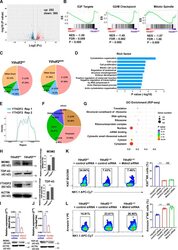
- Figure S5. Transcriptome-wide RNA-seq, m 6 A-seq, and RIP-seq assays in murine NK cells. (A) Volcano plots of differentially expressed genes from RNA-seq. (B) GSEA showing enrichment of E2F targets, G2/M checkpoint, and mitotic spindle hallmark gene sets in Ythdf2 DeltaNK NK cells compared with Ythdf2 WT NK cells. (C) The proportion of m 6 A peak distribution in NK cells from Ythdf2 WT and Ythdf2 DeltaNK mice. (D) GO analysis of transcripts with m 6 A peaks. (E) Density distribution of the YTHDF2-binding sites across mRNA transcriptome from RIP-seq data. (F) The proportion of YTHDF2 binding site distribution from RIP-seq data. (G) Top 10 GO clusters from GO analysis of YTHDF2 target genes from RIP-seq data. (H) Immunoblotting showing the protein levels of MDM2, TDP-43, and YTHDF2 in IL-2-expanded NK cells enriched from the spleen of Ythdf2 WT and Ythdf2 DeltaNK mice. (I and J) qPCR and immunoblotting showing the expression of Mdm2 (I) and Tardbp (J) in NK cells transfected with gene-specific or control siRNA. (K and L) IL-2-expanded NK cells were transfected with Mdm2 -specific siRNA cells under the stimulation of IL-15. 3 d later, cell proliferation and apoptosis were analyzed by Ki67 staining (K) and annexin V staining (L), respectively ( n = 3 per group). Data are shown as mean +- SD and were analyzed by unpaired two-tailed t test (I and J) or one-way ANOVA with Sidak post-test (K and L). Data are representative of at least two independent experiments. ***, P < 0.001. ES,