 Explore
 Validate
 Learn
Learn Western blot
Antibody data
- Antibody Data
- Antigen structure
- References [0]
- Comments [0]
- Validations
- Western blot [3]
- Immunocytochemistry [1]
- Immunohistochemistry [1]
Submit
Validation data
Reference
Comment
Report error
- Product number
- MAB7778 - Provider product page

- Provider
- R&D Systems
- Product name
- Human/Mouse/Rat TDP-43/TARDBP Antibody
- Antibody type
- Monoclonal
- Description
- Protein A or G purified from hybridoma culture supernatant. Detects human TDP-43/TARDBP in direct ELISAs. Detects human, mouse, and rat TDP-43/TARDBP in Western blots.
- Reactivity
- Human, Mouse, Rat
- Host
- Mouse
- Conjugate
- Unconjugated
- Antigen sequence
Q13148- Isotype
- IgG
- Antibody clone number
- 671834
- Vial size
- 100 ug
- Concentration
- LYOPH
- Storage
- Use a manual defrost freezer and avoid repeated freeze-thaw cycles. 12 months from date of receipt, -20 to -70 °C as supplied. 1 month, 2 to 8 °C under sterile conditions after reconstitution. 6 months, -20 to -70 °C under sterile conditions after reconstitution.
No comments: Submit comment
Supportive validation
- Submitted by
- R&D Systems (provider)
- Main image
- Experimental details
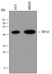
- Detection of Human TDP-43/TARDBP by Western Blot. Western blot shows lysates of A431 human epithelial carcinoma cell line and NR8383 rat alveolar macrophage cell line. PVDF membrane was probed with 0.4 µg/mL of Mouse Anti-Human/Mouse/Rat TDP-43/TARDBP Monoclonal Antibody (Catalog # MAB7778) followed by HRP-conjugated Anti-Mouse IgG Secondary Antibody (Catalog # HAF018). A specific band was detected for TDP-43/TARDBP at approximately 45 kDa (as indicated). This experiment was conducted under reducing conditions and using Immunoblot Buffer Group 1.

- Submitted by
- R&D Systems (provider)
- Main image
- Experimental details
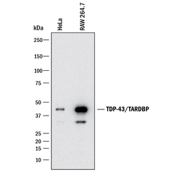
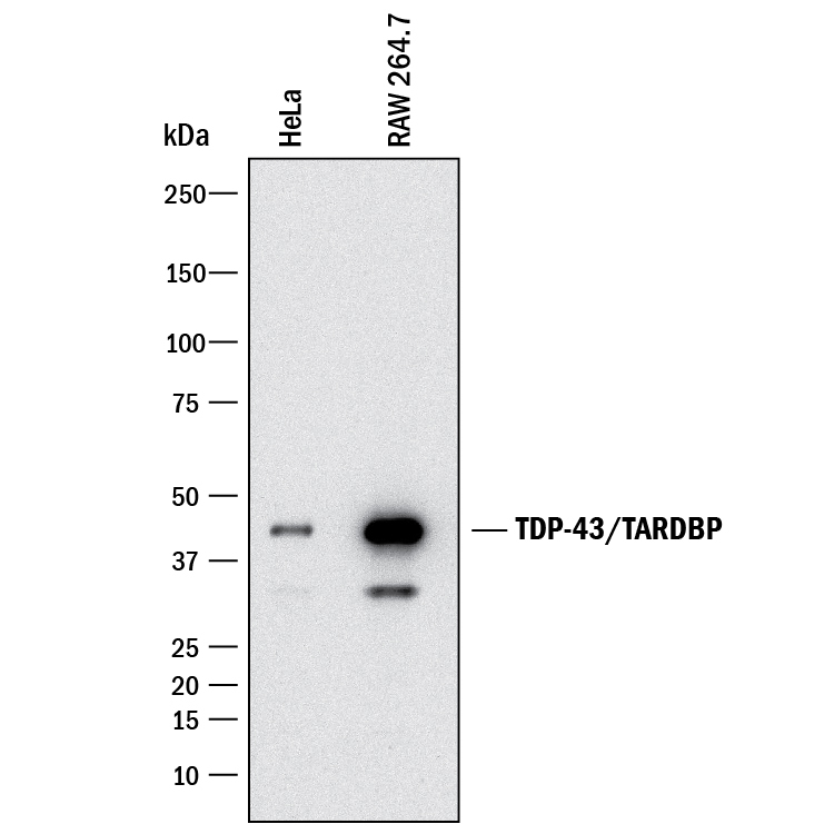
- Detection of Human and Mouse TDP-43/TARDBP by Western Blot. Western blot shows lysates of HeLa human cervical epithelial carcinoma cell line and RAW 264.7 mouse monocyte/macrophage cell line. PVDF membrane was probed with 1 µg/mL of Mouse Anti-Human TDP-43/TARDBP Monoclonal Antibody (Catalog # MAB7778) followed by HRP-conjugated Anti-Mouse IgG Secondary Antibody (Catalog # HAF018). A specific band was detected for TDP-43/TARDBP at approximately 43 kDa (as indicated). This experiment was conducted under reducing conditions and using Immunoblot Buffer Group 3.
- Submitted by
- R&D Systems (provider)
- Main image
- Experimental details
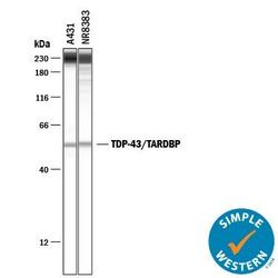
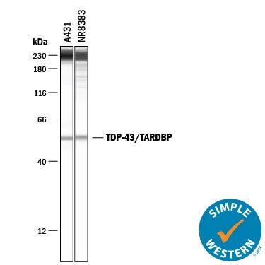
- Detection of Human and Rat TDP-43/TARDBP by Simple WesternTM. Simple Western lane view shows lysates of A431 human epithelial carcinoma cell line and NR8383 rat alveolar macrophage cell line, loaded at 0.2 mg/mL. A specific band was detected for TDP-43/TARDBP at approximately 54 kDa (as indicated) using 5 µg/mL of Mouse Anti-Human TDP-43/TARDBP Monoclonal Antibody (Catalog # MAB7778) . This experiment was conducted under reducing conditions and using the 12-230 kDa separation system. Non-specific interaction with the 230 kDa Simple Western standard may be seen with this antibody.
Supportive validation
- Submitted by
- R&D Systems (provider)
- Main image
- Experimental details
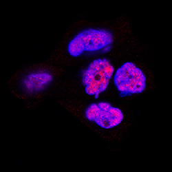
- TDP-43/TARDBP in A431 Human Cell Line. TDP-43/TARDBP was detected in immersion fixed A431 human epithelial carcinoma cell line using Mouse Anti-Human/Mouse/Rat TDP-43/TARDBP Monoclonal Antibody (Catalog # MAB7778) at 1 µg/mL for 3 hours at room temperature. Cells were stained using the NorthernLights™ 557-conjugated Anti-Sheep IgG Secondary Antibody (red; Catalog # NL010) and counterstained with DAPI (blue). Specific staining was localized to nuclei. View our protocol for Fluorescent ICC Staining of Cells on Coverslips.

Supportive validation
- Submitted by
- R&D Systems (provider)
- Main image
- Experimental details
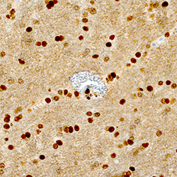
- TDP-43/TARDBP in Human Brain. TDP-43/TARDBP was detected in immersion fixed paraffin-embedded sections of human brain (hippocampus) using Mouse Anti-Human/Mouse/Rat TDP-43/TARDBP Monoclonal Antibody (Catalog # MAB7778) at 1.7 µg/mL overnight at 4 °C. Tissue was stained using the Anti-Mouse HRP-DAB Cell & Tissue Staining Kit (brown; Catalog # CTS002) and counterstained with hematoxylin (blue). Specific staining was localized to nuclei. View our protocol for Chromogenic IHC Staining of Paraffin-embedded Tissue Sections.
