Explore
 Validate
 Learn
Learn Western blot
 Immunocytochemistry
Antibody data
- Antibody Data
- Antigen structure
- References [1]
- Comments [0]
- Validations
- Western blot [3]
- Immunohistochemistry [1]
Submit
Validation data
Reference
Comment
Report error
- Product number
- NBP1-82979 - Provider product page

- Provider
- Novus Biologicals
- Proper citation
- Novus Cat#NBP1-82979, RRID:AB_11038475
- Product name
- Rabbit Polyclonal SWAP70 Antibody
- Antibody type
- Polyclonal
- Description
- Immunogen affinity purified. Specificity of human SWAP70 antibody verified on a Protein Array containing target protein plus 383 other non-specific proteins.
- Reactivity
- Human, Mouse, Rat
- Host
- Rabbit
- Isotype
- IgG
- Vial size
- 0.1 ml
- Storage
- Store at 4C short term. Aliquot and store at -20C long term. Avoid freeze-thaw cycles.
Submitted references SWAP70 Organizes the Actin Cytoskeleton and Is Essential for Phagocytosis.
Baranov MV, Revelo NH, Dingjan I, Maraspini R, Ter Beest M, Honigmann A, van den Bogaart G
Cell reports 2016 Nov 1;17(6):1518-1531
Cell reports 2016 Nov 1;17(6):1518-1531
No comments: Submit comment
Supportive validation
- Submitted by
- Novus Biologicals (provider)
- Main image
- Experimental details
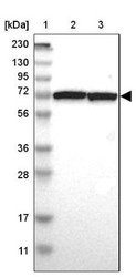
- Western Blot: SWAP70 Antibody [NBP1-82979] - Lane 1: Marker [kDa] 230, 130, 95, 72, 56, 36, 28, 17, 11. Lane 2: Human cell line RT-4. Lane 3: Human cell line U-251MG sp
- Submitted by
- Novus Biologicals (provider)
- Main image

- Experimental details
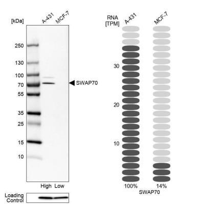
- Western Blot: SWAP70 Antibody [NBP1-82979] - Analysis in human cell lines A-431 and MCF-7. Corresponding RNA-seq data are presented for the same cell lines. Loading control: Anti-PPIB.
- Submitted by
- Novus Biologicals (provider)
- Main image
- Experimental details
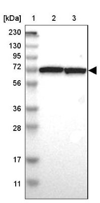
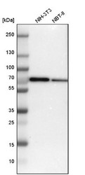
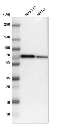
- Western Blot: SWAP70 Antibody [NBP1-82979] - Analysis in mouse cell line NIH-3T3 and rat cell line NBT-II.
Supportive validation
- Submitted by
- Novus Biologicals (provider)
- Main image
- Experimental details
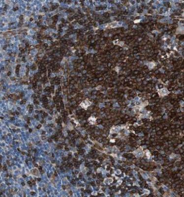
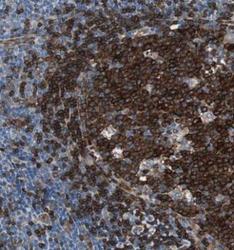
- Immunohistochemistry-Paraffin: SWAP70 Antibody [NBP1-82979] - Staining of human lymph node shows strong cytoplasmic positivity in germinal center cells.