Explore
 Validate
 Learn
Learn Western blot
Antibody data
- Antibody Data
- Antigen structure
- References [0]
- Comments [0]
- Validations
- Western blot [1]
- Immunocytochemistry [3]
- Immunoprecipitation [1]
- Immunohistochemistry [9]
- Other assay [1]
Submit
Validation data
Reference
Comment
Report error
- Product number
- PA5-54183 - Provider product page

- Provider
- Invitrogen Antibodies
- Product name
- Ataxin 2 Polyclonal Antibody
- Antibody type
- Polyclonal
- Antigen
- Recombinant protein fragment
- Description
- Immunogen sequence: EGHSINTREN KYIPPGQRNR EVISWGSGRQ NSPRMGQPGS GSMPSRSTSH TSDFNPNSGS DQRVVNGGVP WPSPCPSPSS RPPSRYQSGP NSLPPRAATP TRPPSRPPSR PSRPPSHPSA HGSPAPVSTM PKRMSSE Highest antigen sequence identity to the following orthologs: Mouse - 93%, Rat - 95%.
- Reactivity
- Human
- Host
- Rabbit
- Isotype
- IgG
- Vial size
- 100 μL
- Concentration
- 0.5 mg/mL
- Storage
- Store at 4°C short term. For long term storage, store at -20°C, avoiding freeze/thaw cycles.
No comments: Submit comment
Supportive validation
- Submitted by
- Invitrogen Antibodies (provider)
- Main image
- Experimental details
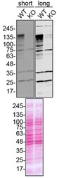
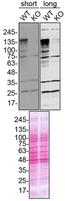
- Western blot analysis of Ataxin-2 was performed by loading 10 µg of WT (lane 1) and ATXN CRISPR KO (lane 2) HAP1 cell lysates in RIPA buffer onto a 4-15% gradient polyacrylamide gel. Proteins were transferred to nitrocellulose membrane and blocked in 5% milk. Ponceau stained transfer of blot is shown. Ataxin-2 was detected using an Ataxin-2 polyclonal antibody (Product # PA5-54183) at a dilution of 1:2,000 in 5% BSA in TBST overnight at 4 deg, followed by secondary antibody diluted to 0.2 µg/mL using Goat anti-Rabbit IgG (H+L) HRP antibody (Product # 65-6120). Chemiluminescent detection was performed using Pierce ECL Western Blotting Substrate (Product # 32106). Data courtesy of YCharOS Inc., an open science company with the mission of characterizing commercially available antibodies using knockout validation.
Supportive validation
- Submitted by
- Invitrogen Antibodies (provider)
- Main image
- Experimental details
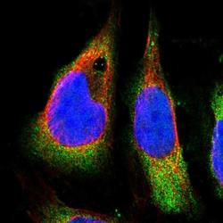
- Immunofluorescent staining of Ataxin 2 in human cell line U-2 OS shows positivity in cytoplasm. Samples were probed using an Ataxin 2 Polyclonal Antibody (Product # PA5-54183).

- Submitted by
- Invitrogen Antibodies (provider)
- Main image

- Experimental details
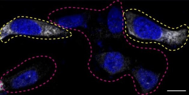
- Immunofluorescence of Ataxin 2 was performed using parental and Ataxin-2 HAP1 CRISPR KO cells that were transfected with a GFP or mCherry plasmid, respectively. At 48 hrs post transfection parental and KO cells were mixed and plated to a 1:1 ratio on coverslips as a mosaic and incubated for 24 hrs. Cells were fixed in 4% PFA for 15 min and permeabilized with 0.1% Triton X-100. Cells were stained with Ataxin polyclonal antibody (Product # PA5-54183) overnight at 4 deg. Secondary antibody incubation was performed using 1 µg/mL of goat anti-rabbit IgG (H+L) Alexa Fluor 555 secondary antibody (Product # A-21429) for 1 hr at RT. Imaging was performed with a 40X oil objective and analysis was performed using Image J. Cell image represents a single focal plane. Data courtesy of YCharOS Inc., an open science company with the mission of characterizing commercially available antibodies using knockout validation.
- Submitted by
- Invitrogen Antibodies (provider)
- Main image
- Experimental details
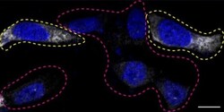
- Immunofluorescence of Ataxin 2 was performed using parental and Ataxin-2 HAP1 CRISPR KO cells that were transfected with a GFP or mCherry plasmid, respectively. At 48 hrs post transfection parental and KO cells were mixed and plated to a 1:1 ratio on coverslips as a mosaic and incubated for 24 hrs. Cells were fixed in 4% PFA for 15 min and permeabilized with 0.1% Triton X-100. Cells were stained with Ataxin polyclonal antibody (Product # PA5-54183) overnight at 4 deg. Secondary antibody incubation was performed using 1 µg/mL of goat anti-rabbit IgG (H+L) Alexa Fluor 555 secondary antibody (Product # A-21429) for 1 hr at RT. Imaging was performed with a 40X oil objective and analysis was performed using Image J. Cell image represents a single focal plane. Data courtesy of YCharOS Inc., an open science company with the mission of characterizing commercially available antibodies using knockout validation.

Supportive validation
- Submitted by
- Invitrogen Antibodies (provider)
- Main image

- Experimental details
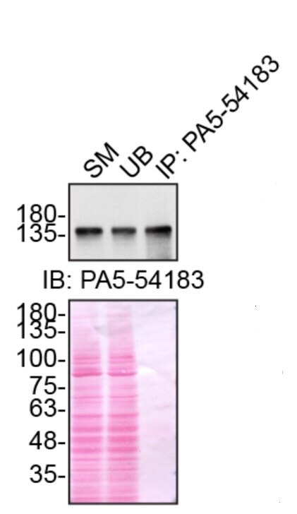
- Immunoprecipitation of Ataxin was performed on HAP1 cell lysates. Antibody-bead conjugates were prepared by adding 1 µg of Ataxin polyclonal antibody (Product # PA5-54183) with 30 µL of protein A -Sepharose beads and rocked overnight at 4 deg. 1 µg of lysate was incubated with antibody-bead conjugate for 2 hrs at 4 deg. After multiple washes, 10% starting material (SM), 10% unbound fraction (UB) and immunoprecipitated fraction (IP) were processed for immunoblot using the same ATXN antibody (Product # PA5-54183). Ponceau stained transfer of blot is shown. Data courtesy of YCharOS Inc., an open science company with the mission of characterizing commercially available antibodies using knockout validation.

Supportive validation
- Submitted by
- Invitrogen Antibodies (provider)
- Main image
- Experimental details
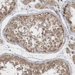
- Immunohistochemical staining of Ataxin 2 in human testis using Ataxin 2 Polyclonal Antibody (Product # PA5-54183) shows moderate to strong cytoplasmic positivity in cells in seminiferous ducts.

- Submitted by
- Invitrogen Antibodies (provider)
- Main image
- Experimental details
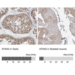
- Immunohistochemical staining of Ataxin 2 in human testis and skeletal muscle tissues using Ataxin 2 Polyclonal Antibody (Product # PA5-54183). Corresponding ATXN2 RNA-seq data are presented for the same tissues.

- Submitted by
- Invitrogen Antibodies (provider)
- Main image

- Experimental details
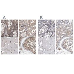
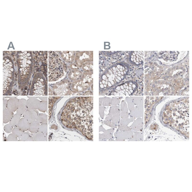
- Immunohistochemical staining of Ataxin 2 in human colon, kidney, skeletal muscle and testis using Ataxin 2 Polyclonal Antibody (Product # PA5-54183) (A) shows similar protein distribution across tissues to an independent Ataxin 2 Polyclonal Antibody (B).

- Submitted by
- Invitrogen Antibodies (provider)
- Main image
- Experimental details
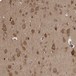
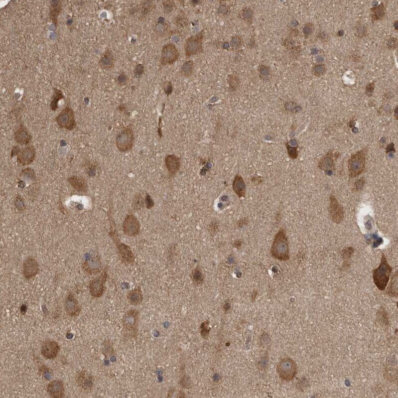
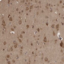
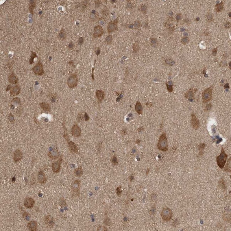
- Immunohistochemical staining of Ataxin 2 in human cerebral cortex using Ataxin 2 Polyclonal Antibody (Product # PA5-54183) shows moderate to strong cytoplasmic positivity in neurons.
- Submitted by
- Invitrogen Antibodies (provider)
- Main image
- Experimental details
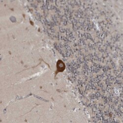
- Immunohistochemical staining of Ataxin 2 in human cerebellum using Ataxin 2 Polyclonal Antibody (Product # PA5-54183) shows moderate to strong cytoplasmic positivity in purkinje cells.

- Submitted by
- Invitrogen Antibodies (provider)
- Main image

- Experimental details
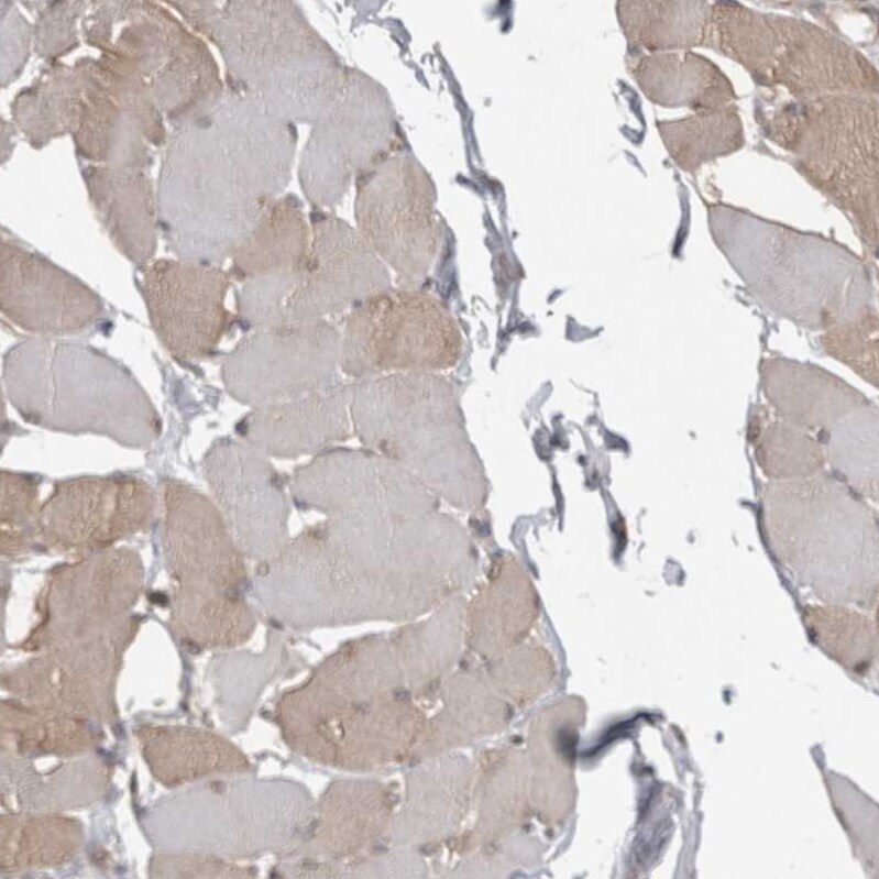
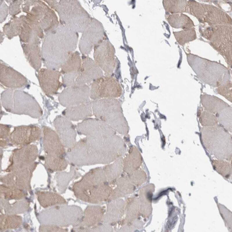
- Immunohistochemical staining of Ataxin 2 in human skeletal muscle using Ataxin 2 Polyclonal Antibody (Product # PA5-54183) shows weak to moderate cytoplasmic positivity in a subset of myocytes.
- Submitted by
- Invitrogen Antibodies (provider)
- Main image
- Experimental details
- Immunohistochemical staining of Ataxin 2 in human colon, kidney, skeletal muscle and testis using Ataxin 2 Polyclonal Antibody (Product # PA5-54183) (A) shows similar protein distribution across tissues to an independent Ataxin 2 Polyclonal Antibody (B).
- Submitted by
- Invitrogen Antibodies (provider)
- Main image
- Experimental details
- Immunohistochemical staining of Ataxin 2 in human cerebral cortex using Ataxin 2 Polyclonal Antibody (Product # PA5-54183) shows moderate to strong cytoplasmic positivity in neurons.
- Submitted by
- Invitrogen Antibodies (provider)
- Main image

- Experimental details
- Immunohistochemical staining of Ataxin 2 in human skeletal muscle using Ataxin 2 Polyclonal Antibody (Product # PA5-54183) shows weak to moderate cytoplasmic positivity in a subset of myocytes.
Supportive validation
- Submitted by
- Invitrogen Antibodies (provider)
- Main image

- Experimental details
- Immunoprecipitation of Ataxin was performed on HAP1 cell lysates. Antibody-bead conjugates were prepared by adding 1 µg of Ataxin polyclonal antibody (Product # PA5-54183) with 30 µL of protein A -Sepharose beads and rocked overnight at 4 deg. 1 µg of lysate was incubated with antibody-bead conjugate for 2 hrs at 4 deg. After multiple washes, 10% starting material (SM), 10% unbound fraction (UB) and immunoprecipitated fraction (IP) were processed for immunoblot using the same ATXN antibody (Product # PA5-54183). Ponceau stained transfer of blot is shown. Data courtesy of YCharOS Inc., an open science company with the mission of characterizing commercially available antibodies using knockout validation.