Explore
 Validate
 Learn
Learn Western blot
 Immunocytochemistry
Antibody data
- Antibody Data
- Antigen structure
- References [1]
- Comments [0]
- Validations
- Immunocytochemistry [8]
- Immunohistochemistry [6]
- Flow cytometry [7]
- Other assay [2]
Submit
Validation data
Reference
Comment
Report error
- Product number
- PA5-78845 - Provider product page

- Provider
- Invitrogen Antibodies
- Product name
- Ataxin 2 Polyclonal Antibody
- Antibody type
- Polyclonal
- Antigen
- Synthetic peptide
- Description
- Reconstitute with 0.2 mL of distilled water to yield a concentration of 500 µg/mL. Positive Control - WB: human K562 whole cell, human A549 whole cell, rat brain tissue, rat liver tissue, mouse brain tissue, mouse liver tissue. IHC: human breast cancer tissue, human rectal cancer tissue, mouse brain tissue, rat brain tissue. Flow: RT4 cell.
- Reactivity
- Human, Mouse, Rat
- Host
- Rabbit
- Isotype
- IgG
- Vial size
- 100 μg
- Concentration
- 500 μg/mL
- Storage
- -20°C
Submitted references Intracellular dynamics of Ataxin-2 in the human brains with normal and frontotemporal lobar degeneration with TDP-43 inclusions.
Watanabe R, Higashi S, Nonaka T, Kawakami I, Oshima K, Niizato K, Akiyama H, Yoshida M, Hasegawa M, Arai T
Acta neuropathologica communications 2020 Oct 28;8(1):176
Acta neuropathologica communications 2020 Oct 28;8(1):176
No comments: Submit comment
Supportive validation
- Submitted by
- Invitrogen Antibodies (provider)
- Main image
- Experimental details
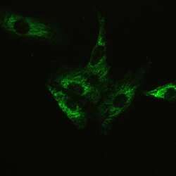
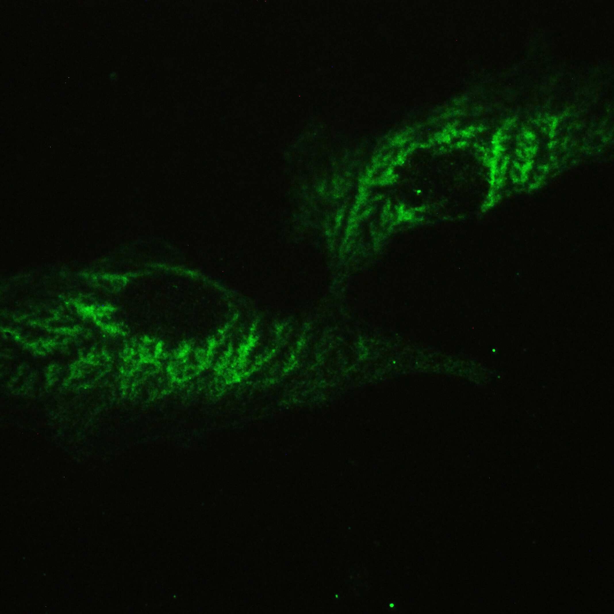
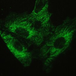
- Immunofluorescent analysis of Ataxin 2 on A549 cells. Antigen retrieval was performed using citrate buffer (pH6, epitope retrieval solution) for 20 mins. Sample was blocked using 10% goat serum, incubated with Ataxin 2 polyclonal antibody (Product# PA5-78845) with a dilution of 2 µg/mL (overnight at 4°C). Development was performed using Streptavidin-Biotin-Complex (SABC) with DAB chromogen method.

- Submitted by
- Invitrogen Antibodies (provider)
- Main image

- Experimental details
- Immunofluorescent analysis of Ataxin 2 on A549 cells. Antigen retrieval was performed using citrate buffer (pH6, epitope retrieval solution) for 20 mins. Sample was blocked using 10% goat serum, incubated with Ataxin 2 polyclonal antibody (Product# PA5-78845) with a dilution of 2 µg/mL (overnight at 4°C). Development was performed using Streptavidin-Biotin-Complex (SABC) with DAB chromogen method.

- Submitted by
- Invitrogen Antibodies (provider)
- Main image

- Experimental details
- Immunofluorescent analysis of Ataxin 2 on A549 cells. Antigen retrieval was performed using citrate buffer (pH6, epitope retrieval solution) for 20 mins. Sample was blocked using 10% goat serum, incubated with Ataxin 2 polyclonal antibody (Product# PA5-78845) with a dilution of 2 µg/mL (overnight at 4°C). Development was performed using Streptavidin-Biotin-Complex (SABC) with DAB chromogen method.
Supportive validation
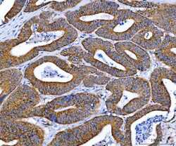
- Submitted by
- Invitrogen Antibodies (provider)
- Main image
- Experimental details
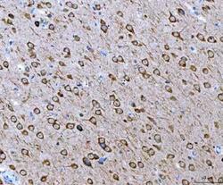
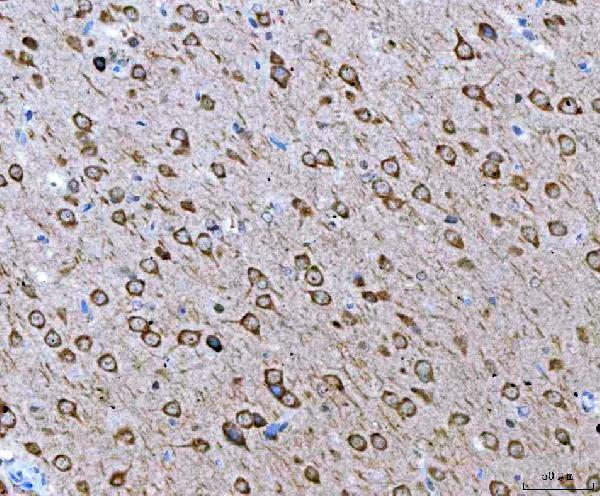
- Immunohistochemistry (Paraffin) analysis of Ataxin 2 in paraffin-embedded section of rat brain tissue using Ataxin 2 Polyclonal Antibody (Product # PA5-78845). Heat mediated antigen retrieval was performed in EDTA buffer (pH 8.0, epitope retrieval solution). The tissue section was blocked with 10% goat serum. The tissue section was then incubated with the primary antibody at a 2 µg/mL dilution overnight at 4°C. Peroxidase conjugated goat anti-rabbit IgG was used as secondary antibody and incubated for 30 minutes at 37°C. The tissue section was developed using HRP Conjugated Rabbit IgG Super Vision Assay Kit with DAB as the chromogen.
- Submitted by
- Invitrogen Antibodies (provider)
- Main image
- Experimental details
- Immunohistochemistry (Paraffin) analysis of Ataxin 2 in paraffin-embedded section of mouse brain tissue using Ataxin 2 Polyclonal Antibody (Product # PA5-78845). Heat mediated antigen retrieval was performed in EDTA buffer (pH 8.0, epitope retrieval solution). The tissue section was blocked with 10% goat serum. The tissue section was then incubated with the primary antibody at a 2 µg/mL dilution overnight at 4°C. Peroxidase conjugated goat anti-rabbit IgG was used as secondary antibody and incubated for 30 minutes at 37°C. The tissue section was developed using HRP Conjugated Rabbit IgG Super Vision Assay Kit with DAB as the chromogen.

- Submitted by
- Invitrogen Antibodies (provider)
- Main image
- Experimental details
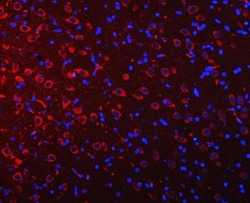
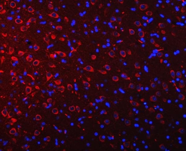
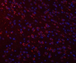
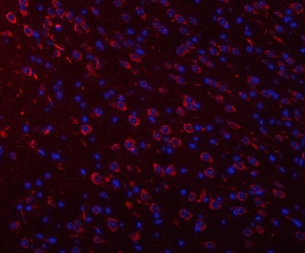
- Immunohistochemistry (Paraffin) analysis of Ataxin 2 in paraffin-embedded section of rat brain tissue using Ataxin 2 Polyclonal Antibody (Product # PA5-78845). Heat mediated antigen retrieval was performed in EDTA buffer (pH 8.0, epitope retrieval solution). The tissue section was blocked with 10% goat serum. The tissue section was then incubated with the primary antibody at a 5 µg/mL dilution overnight at 4°C. Cy3 conjugated goat anti-rabbit IgG was used as secondary antibody at 1:500 dilution and incubated for 30 minutes at 37°C. The section was counterstained with DAPI. Visualize using a fluorescence microscope and filter sets appropriate for the label used.
- Submitted by
- Invitrogen Antibodies (provider)
- Main image
- Experimental details
- Immunohistochemistry (Paraffin) analysis of Ataxin 2 in paraffin-embedded section of mouse brain tissue using Ataxin 2 Polyclonal Antibody (Product # PA5-78845). Heat mediated antigen retrieval was performed in EDTA buffer (pH 8.0, epitope retrieval solution). The tissue section was blocked with 10% goat serum. The tissue section was then incubated with the primary antibody at a 5 µg/mL dilution overnight at 4°C. Cy3 conjugated goat anti-rabbit IgG was used as secondary antibody at 1:500 dilution and incubated for 30 minutes at 37°C. The section was counterstained with DAPI. Visualize using a fluorescence microscope and filter sets appropriate for the label used.
- Submitted by
- Invitrogen Antibodies (provider)
- Main image
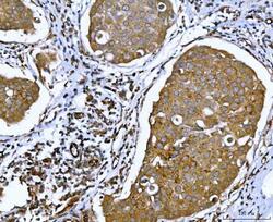
- Experimental details
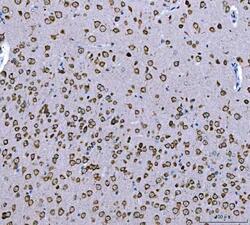
- Immunohistochemistry (Paraffin) analysis of Ataxin 2 in paraffin-embedded section of human breast cancer tissue using Ataxin 2 Polyclonal Antibody (Product # PA5-78845). Heat mediated antigen retrieval was performed in EDTA buffer (pH 8.0, epitope retrieval solution). The tissue section was blocked with 10% goat serum. The tissue section was then incubated with the primary antibody at a 2 µg/mL dilution overnight at 4°C. Peroxidase conjugated goat anti-rabbit IgG was used as secondary antibody and incubated for 30 minutes at 37°C. The tissue section was developed using HRP Conjugated Rabbit IgG Super Vision Assay Kit with DAB as the chromogen.
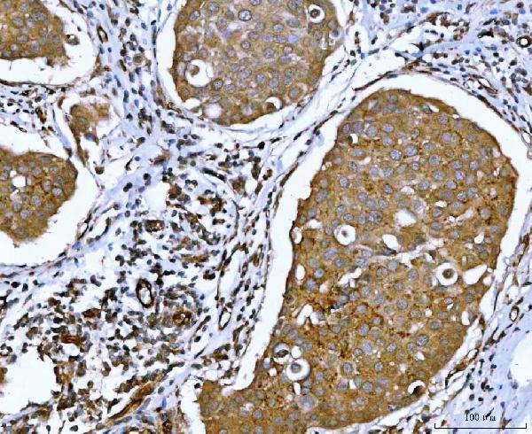
- Submitted by
- Invitrogen Antibodies (provider)
- Main image
- Experimental details
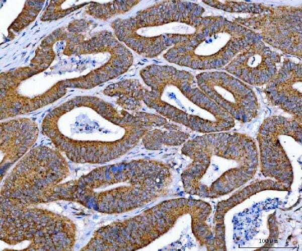
- Immunohistochemistry (Paraffin) analysis of Ataxin 2 in paraffin-embedded section of human rectal cancer tissue using Ataxin 2 Polyclonal Antibody (Product # PA5-78845). Heat mediated antigen retrieval was performed in EDTA buffer (pH 8.0, epitope retrieval solution). The tissue section was blocked with 10% goat serum. The tissue section was then incubated with the primary antibody at a 2 µg/mL dilution overnight at 4°C. Peroxidase conjugated goat anti-rabbit IgG was used as secondary antibody and incubated for 30 minutes at 37°C. The tissue section was developed using HRP Conjugated Rabbit IgG Super Vision Assay Kit with DAB as the chromogen.
Supportive validation
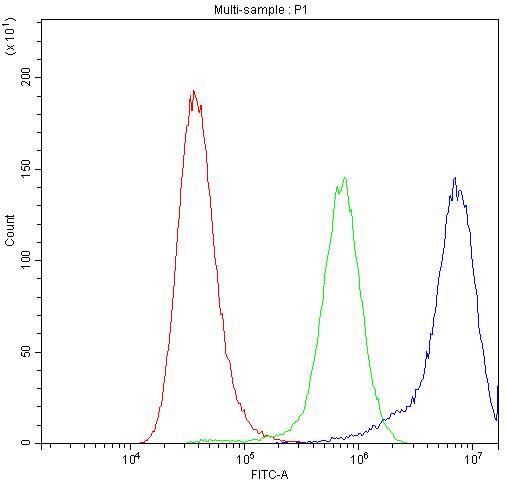
- Submitted by
- Invitrogen Antibodies (provider)
- Main image

- Experimental details
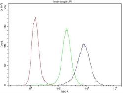
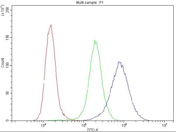
- Flow Cytometry of Ataxin 2 in A549 cells (blue line), isotype control rabbit IgG (green line) and unlabeled (red line). Samples were blocked with 10% goat serum, incubated with Ataxin 2 Polyclonal Antibody (Product # PA5-78845) at a dilution of 1 μg (per 1x10^6 cells), followed by DyLight®488 conjugated goat anti-rabbit IgG (for 30 minutes at 20°C) using 5-10 μg (per 1x10^6 cells) dilution.

- Submitted by
- Invitrogen Antibodies (provider)
- Main image
- Experimental details
- Flow Cytometry of Ataxin 2 in A431 cells (blue line), isotype control rabbit IgG (green line) and unlabeled (red line). Samples were blocked with 10% goat serum, incubated with Ataxin 2 Polyclonal Antibody (Product # PA5-78845) at a dilution of 1 μg (per 1x10^6 cells), followed by DyLight®488 conjugated goat anti-rabbit IgG (for 30 minutes at 20°C) using 5-10 μg (per 1x10^6 cells) dilution.

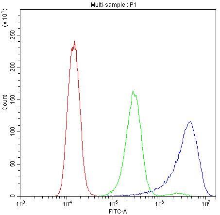
- Submitted by
- Invitrogen Antibodies (provider)
- Main image

- Experimental details
- Flow Cytometry of Ataxin 2 in K562 cells (blue line), isotype control rabbit IgG (green line) and unlabeled (red line). Samples were blocked with 10% goat serum, incubated with Ataxin 2 Polyclonal Antibody (Product # PA5-78845) at a dilution of 1 μg (per 1x10^6 cells), followed by DyLight®488 conjugated goat anti-rabbit IgG (for 30 minutes at 20°C) using 5-10 μg (per 1x10^6 cells) dilution.

- Submitted by
- Invitrogen Antibodies (provider)
- Main image
- Experimental details
- Flow Cytometry of Ataxin 2 in K562 cells (blue line), isotype control rabbit IgG (green line) and unlabeled (red line). Samples were blocked with 10% goat serum, incubated with Ataxin 2 Polyclonal Antibody (Product # PA5-78845) at a dilution of 1 μg (per 1x10^6 cells), followed by DyLight®488 conjugated goat anti-rabbit IgG (for 30 minutes at 20°C) using 5-10 μg (per 1x10^6 cells) dilution.
- Submitted by
- Invitrogen Antibodies (provider)
- Main image

- Experimental details
- Flow Cytometry of Ataxin 2 in A549 cells (blue line), isotype control rabbit IgG (green line) and unlabeled (red line). Samples were blocked with 10% goat serum, incubated with Ataxin 2 Polyclonal Antibody (Product # PA5-78845) at a dilution of 1 μg (per 1x10^6 cells), followed by DyLight®488 conjugated goat anti-rabbit IgG (for 30 minutes at 20°C) using 5-10 μg (per 1x10^6 cells) dilution.
- Submitted by
- Invitrogen Antibodies (provider)
- Main image

- Experimental details
- Flow Cytometry of Ataxin 2 in A431 cells (blue line), isotype control rabbit IgG (green line) and unlabeled (red line). Samples were blocked with 10% goat serum, incubated with Ataxin 2 Polyclonal Antibody (Product # PA5-78845) at a dilution of 1 μg (per 1x10^6 cells), followed by DyLight®488 conjugated goat anti-rabbit IgG (for 30 minutes at 20°C) using 5-10 μg (per 1x10^6 cells) dilution.
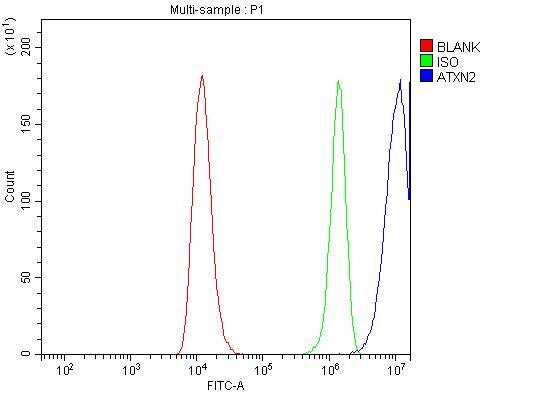
- Submitted by
- Invitrogen Antibodies (provider)
- Main image
- Experimental details
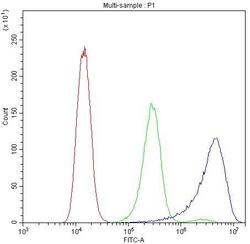
- Flow cytometry analysis of Ataxin 2 in RT4 cells using Ataxin 2 Polyclonal Antibody (Product # PA5-78845), shown in overlay histogram (blue line). To facilitate intracellular staining, cells were fixed with 4% paraformaldehyde and permeabilized with permeabilization buffer. The cells were blocked with 10% normal goat serum, and incubated with the primary antibody (1 μg/1x10^6 cells) for 30 min at 20°C. DyLight 488 conjugated goat anti-rabbit IgG (5-10 µg/1x10^6 cells) was used as secondary antibody for 30 minutes at 20°C. Isotype control antibody (Green line) was rabbit IgG (1 µg/1x10^6) used under the same conditions. Unlabelled sample (Red line) was also used as a control.
Supportive validation
- Submitted by
- Invitrogen Antibodies (provider)
- Main image
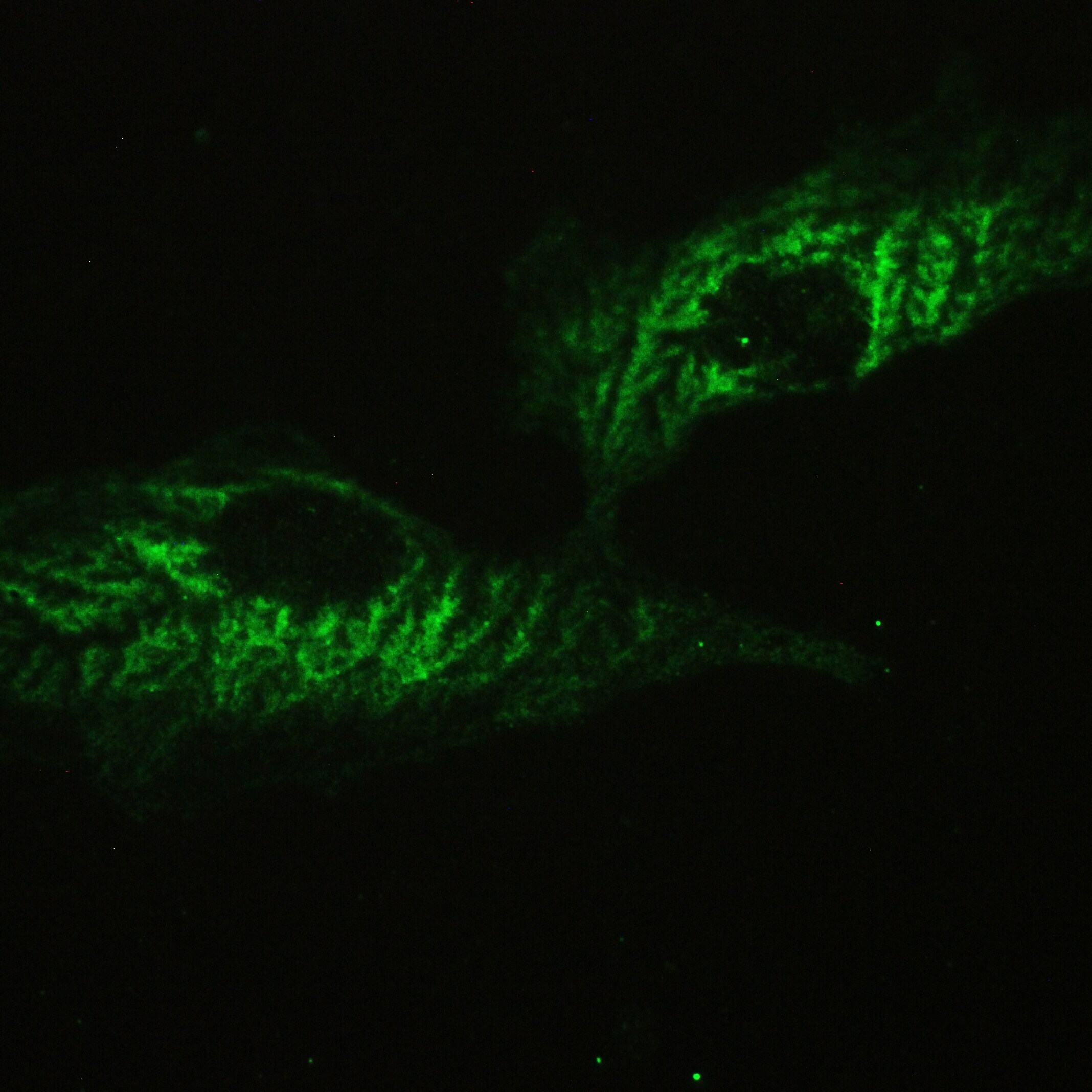
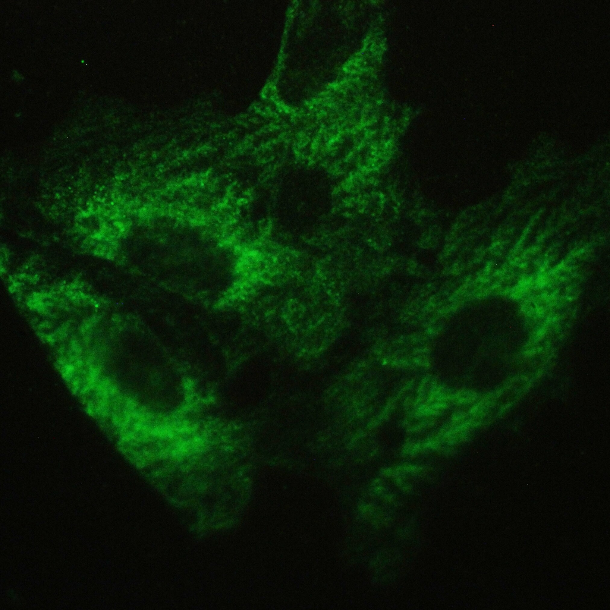
- Experimental details
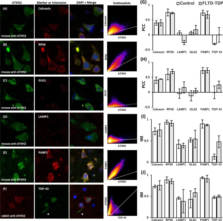
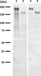
- Fig. 1 Verification of ATXN2 antibodies. Two different ATXN2 antibodies were used in this study. Their specificities were confirmed by western blotting of cultured cells transfected with ATXN2 expression plasmids, pFN21A-Halo-ATXN2 (lane 1, 3) and pcDNA3-Flag-ATXN2 (lane 2, 4). The left blot (lane 1, 2) and the right one (lane 3, 4) were incubated with mouse monoclonal antibody (BD Biosciences, aa713-904) and rabbit polyclonal antibody (Thermofisher, aa1293-1313), respectively. Halo- and Flag-tagged ATXN2 were electrophoresed at approximately 220 kDa and 160 kDa, respectively

- Submitted by
- Invitrogen Antibodies (provider)
- Main image
- Experimental details
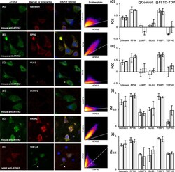
- Fig. 4 Comparison of ATXN2 localization in the neuronal cytoplasm in control and FTLD-TDP brains. a - f The confocal microscopic images of neuronal cytoplasmic ATXN2 and organelle marker proteins or ATXN2's related proteins from a FTLD-TDP patient's brain were obtained as were those of controls. Representative fluorescent double-stained images from the upper layer of the cerebral temporal cortex, including merged images with DAPI staining and two-dimensional scatterplots, are presented. The localization of ATXN2 in the neuronal cytoplasm did not differ from that in the controls shown in Fig. 3 . Note that the LAMP1-immunoreactivity was strikingly decreased ( d ). f In the double-fluorescent images using the rabbit polyclonal anti-ATXN2 antibody and the mouse monoclonal phosphorylation-independent anti-TDP-43 antibody, asterisks indicate that ATXN2 did not appear in TDP-43 positive dystrophic neurites. Pseudo coloring was used for each channel. g - j Colocalization coefficient values for the upper layer of the temporal cortex ( g , i ) and hippocampal CA1 ( h , j ) were obtained from normal controls and FTLD-TDP patients. The mean values of Pearson's correlation coefficients above threshold (PCC) ( g , h ) and Manders' colocalization coefficients above threshold (tM) ( i , j ) are plotted. ATXN2 and Calnexin did not show significant PCC rates but moderate tM rates, indicating their fractional overlapping. RPS6 and PABP1 showed both significant PCC rates and high tM rates with