Explore
 Validate
 Learn
Learn Western blot
 Immunohistochemistry
Antibody data
- Antibody Data
- Antigen structure
- References [1]
- Comments [0]
- Validations
- Western blot [1]
- Immunocytochemistry [2]
- Immunohistochemistry [1]
Submit
Validation data
Reference
Comment
Report error
- Product number
- HPA018295 - Provider product page

- Provider
- Atlas Antibodies
- Proper citation
- Atlas Antibodies Cat#HPA018295, RRID:AB_1845130
- Product name
- Anti-ATXN2
- Antibody type
- Polyclonal
- Description
- Polyclonal Antibody against Human ATXN2, Gene description: ataxin 2, Alternative Gene Names: ATX2, SCA2, TNRC13, Validated applications: IHC, ICC, Uniprot ID: Q99700, Storage: Store at +4°C for short term storage. Long time storage is recommended at -20°C.
- Reactivity
- Human
- Host
- Rabbit
- Conjugate
- Unconjugated
- Isotype
- IgG
- Vial size
- 100 µl
- Concentration
- 0.2 mg/ml
- Storage
- Store at +4°C for short term storage. Long time storage is recommended at -20°C.
- Handling
- The antibody solution should be gently mixed before use.
Submitted references Lateral olfactory tract usher substance (LOTUS), an endogenous Nogo receptor antagonist, ameliorates disease progression in amyotrophic lateral sclerosis model mice.
Ikeda T, Takahashi K, Higashi M, Komiya H, Asano T, Ogasawara A, Kubota S, Hashiguchi S, Kunii M, Tanaka K, Tada M, Doi H, Takeuchi H, Takei K, Tanaka F
Cell death discovery 2023 Dec 14;9(1):454
Cell death discovery 2023 Dec 14;9(1):454
No comments: Submit comment
Enhanced validation
- Submitted by
- klas2
- Enhanced method
- Genetic validation
- Main image
- Experimental details
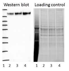
- Western blot of cell lysate from U-2 OS cells transfected with either siRNA targeting ATXN2 or control siRNA. Lane 1: Marker (250, 130, 95, 72, 55, 36, 28, 17, 10) Lane 2: Cell lysate from U-2OS cells transfected with siRNA targeting ATXN2 Lane 3: N/A Lane 4: Cell lysate from U-2OS cells transfected with control siRNA Right image, lane 1-4: loading control
- Sample type
- U-2 OS
- Primary Ab dilution
- 1:275
- Conjugate
- Horseradish Peroxidase
- Secondary Ab
- Secondary Ab
- Secondary Ab dilution
- 1:3000
- Knockdown/Genetic Approaches Application
- Western blot
Enhanced validation
Supportive validation
- Submitted by
- 55af80e3e0991
- Enhanced method
- Genetic validation
- Main image
- Experimental details
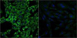
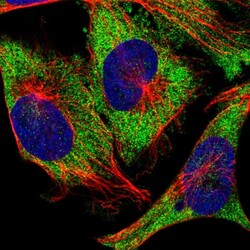
- Confocal images of immunofluorescently stained human U-2 OS cells.The protein ATXN2 is shown in green and the nucleus in blue. The image to the left show cells transfected with control siRNA and the image to the right show cells where ATXN2 has been downregulated with specific siRNA.
- Sample type
- U-2 OS cells
- Primary Ab dilution
- 1:123
- Secondary Ab
- Secondary Ab
- Secondary Ab dilution
- 1:800
- Knockdown/Genetic Approaches Application
- Immunocytochemistry

Supportive validation
- Submitted by
- Atlas Antibodies (provider)
- Main image
- Experimental details
- Immunofluorescent staining of human cell line U-251 MG shows localization to cytosol.
- Sample type
- Human

Supportive validation
- Submitted by
- Atlas Antibodies (provider)
- Enhanced method
- Orthogonal validation
- Main image
- Experimental details
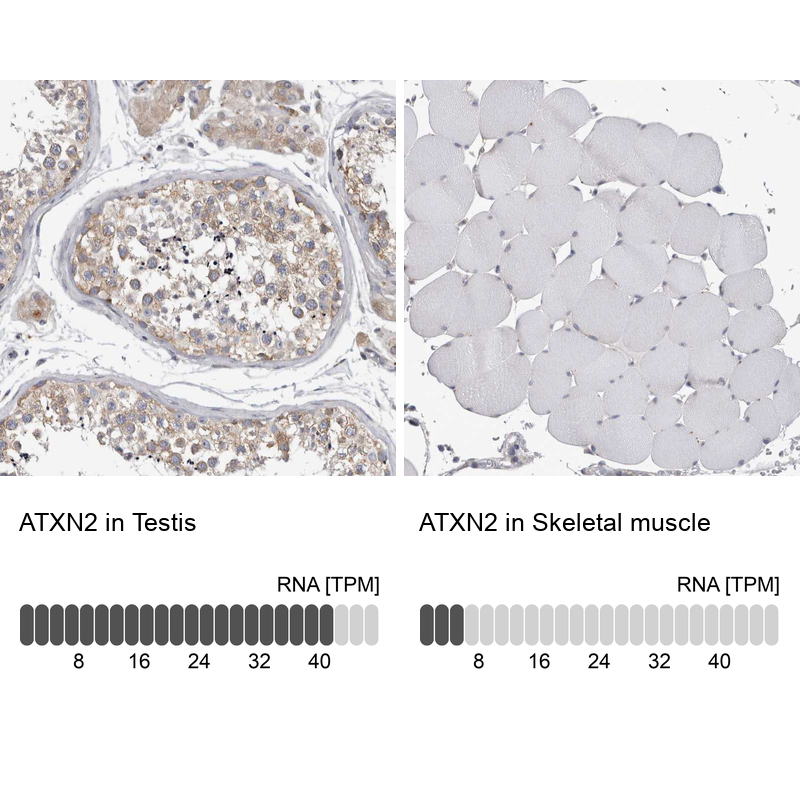
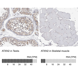
- Immunohistochemistry analysis in human testis and skeletal muscle tissues using HPA018295 antibody. Corresponding ATXN2 RNA-seq data are presented for the same tissues.
- Sample type
- Human
- Protocol
- Protocol