Explore
 Validate
 Learn
Learn Western blot
 Immunoprecipitation
Antibody data
- Antibody Data
- Antigen structure
- References [7]
- Comments [0]
- Validations
- Western blot [2]
- Immunocytochemistry [4]
- Immunohistochemistry [1]
- Other assay [2]
Submit
Validation data
Reference
Comment
Report error
- Product number
- MA1-12640 - Provider product page

- Provider
- Invitrogen Antibodies
- Product name
- XRCC1 Monoclonal Antibody (144)
- Antibody type
- Monoclonal
- Antigen
- Recombinant full-length protein
- Description
- A suggested positive control for this product is HT29, LS174T, HeLa cells or normal testis.
- Reactivity
- Human
- Host
- Mouse
- Isotype
- IgG
- Antibody clone number
- 144
- Vial size
- 100 μL
- Concentration
- 0.2 mg/mL
- Storage
- 4°C
Submitted references Establishment of Acquired Cisplatin Resistance in Ovarian Cancer Cell Lines Characterized by Enriched Metastatic Properties with Increased Twist Expression.
Cadmium treatment suppresses DNA polymerase δ catalytic subunit gene expression by acting on the p53 and Sp1 regulatory axis.
ATM prevents DSB formation by coordinating SSB repair and cell cycle progression.
Cells deficient in base-excision repair reveal cancer hallmarks originating from adjustments to genetic instability.
Association between polymorphisms of XRCC1, p53 and MDR1 genes, the expression of their protein products and prognostic significance in human breast cancer.
Association between polymorphisms of XRCC1, p53 and MDR1 genes, the expression of their protein products and prognostic significance in human breast cancer.
Association of XRCC1 and tyrosyl DNA phosphodiesterase (Tdp1) for the repair of topoisomerase I-mediated DNA lesions.
Bahar E, Kim JY, Kim HS, Yoon H
International journal of molecular sciences 2020 Oct 15;21(20)
International journal of molecular sciences 2020 Oct 15;21(20)
Cadmium treatment suppresses DNA polymerase δ catalytic subunit gene expression by acting on the p53 and Sp1 regulatory axis.
Antoniali G, Marcuzzi F, Casarano E, Tell G
DNA repair 2015 Nov;35:90-105
DNA repair 2015 Nov;35:90-105
ATM prevents DSB formation by coordinating SSB repair and cell cycle progression.
Khoronenkova SV, Dianov GL
Proceedings of the National Academy of Sciences of the United States of America 2015 Mar 31;112(13):3997-4002
Proceedings of the National Academy of Sciences of the United States of America 2015 Mar 31;112(13):3997-4002
Cells deficient in base-excision repair reveal cancer hallmarks originating from adjustments to genetic instability.
Markkanen E, Fischer R, Ledentcova M, Kessler BM, Dianov GL
Nucleic acids research 2015 Apr 20;43(7):3667-79
Nucleic acids research 2015 Apr 20;43(7):3667-79
Association between polymorphisms of XRCC1, p53 and MDR1 genes, the expression of their protein products and prognostic significance in human breast cancer.
Rybárová S, Vecanová J, Hodorová I, Mihalik J, Čižmáriková M, Mojžiš J, Solár P, Benický M, Adamkov M, Mirossay L
Medical science monitor : international medical journal of experimental and clinical research 2011 Dec;17(12):BR354-63
Medical science monitor : international medical journal of experimental and clinical research 2011 Dec;17(12):BR354-63
Association of XRCC1 and tyrosyl DNA phosphodiesterase (Tdp1) for the repair of topoisomerase I-mediated DNA lesions.
Plo I, Liao ZY, Barceló JM, Kohlhagen G, Caldecott KW, Weinfeld M, Pommier Y
DNA repair 2003 Oct 7;2(10):1087-100
DNA repair 2003 Oct 7;2(10):1087-100
No comments: Submit comment
Supportive validation
- Submitted by
- Invitrogen Antibodies (provider)
- Main image
- Experimental details
- Western blot of XRCC1 using XRCC1 Monoclonal Antibody (Product # MA5-12071) on LS174T Cells.
- Submitted by
- Invitrogen Antibodies (provider)
- Main image
- Experimental details
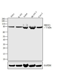
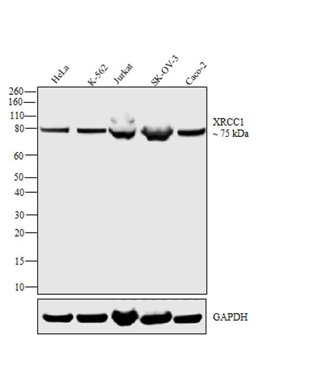
- Western blot analysis was performed on whole cell extract (30 µg lysate) of HeLa (Lane 1), K-562 (Lane 2), Jurkat (Lane 3), SK-OV-3 and Caco-2 (Lane 4). The blot was probed with Mouse Anti-XRCC1 Monoclonal Antibody (Product # MA5-12071, 1 µg/mL) and detected by chemiluminescence using Goat anti-Mouse IgG (H+L) Superclonal™ Secondary Antibody, HRP conjugate (Product # A28177, 0.25 µg/mL, 1:4000 dilution). A 75 kDa band corresponding to XRCC1 was observed across cell lines tested. Known quantity of protein samples were electrophoresed using Novex® NuPAGE® 4-12 % Bis-Tris gel (Product # NP0321BOX), XCell SureLock™ Electrophoresis System (Product # EI0002) and Novex® Sharp Pre-Stained Protein Standard (Product # LC5800). Resolved proteins were then transferred onto a nitrocellulose membrane with iBlot® 2 Dry Blotting System (Product # IB21001). The membrane was probed with the relevant primary and secondary Antibody following blocking with 5 % skimmed milk. Chemiluminescent detection was performed using Pierce™ ECL Western Blotting Substrate (Product # 32106).
Supportive validation
- Submitted by
- Invitrogen Antibodies (provider)
- Main image

- Experimental details
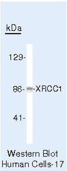
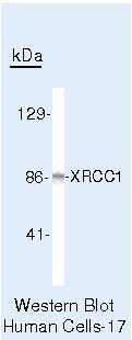
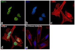
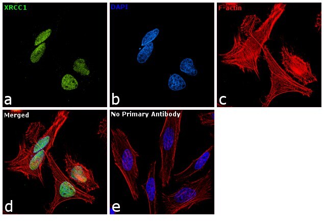
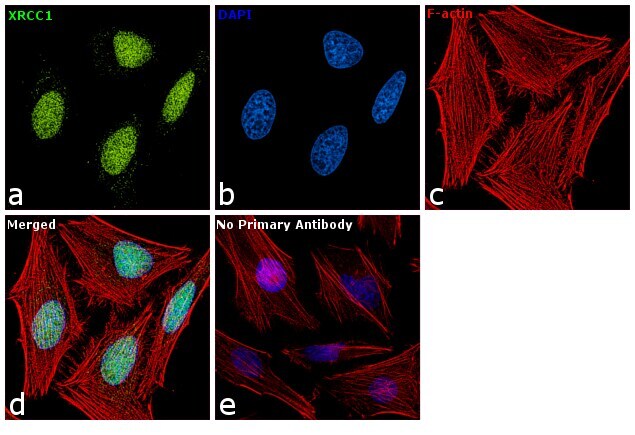
- Immunofluorescence analysis of XRCC1 was performed using 70 % confluent log phase HeLa cells. The cells were fixed with 4% paraformaldehyde for 10 minutes, permeabilized with 0.1% Triton™ X-100 for 10 minutes, and blocked with 1% BSA for 1 hour at room temperature. The cells were labeled with XRCC1 Mouse Monoclonal antibody (Product # MA1-12640) at 2 µg/mL in 0.1% BSA and incubated for 3 hours at room temperature and then labeled with Goat anti-Mouse IgG (H+L) Superclonal™ Secondary Antibody, Alexa Fluor® 488 conjugate (Product # A28175) at a dilution of 1:2000 for 45 minutes at room temperature (Panel a: green). Nuclei (Panel b: blue) were stained with SlowFade® Gold Antifade Mountant with DAPI (S36938). F-actin (Panel c: red) was stained with Rhodamine Phalloidin (Product # R415, 1:300). Panel d represents the merged image showing Nuclear localization. Panel e shows the no primary antibody control. The images were captured at 60X magnification.

- Submitted by
- Invitrogen Antibodies (provider)
- Main image
- Experimental details
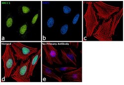
- Immunofluorescence analysis of XRCC1 was performed using 70 % confluent log phase HeLa cells. The cells were fixed with 4% paraformaldehyde for 10 minutes, permeabilized with 0.1% Triton™ X-100 for 10 minutes, and blocked with 1% BSA for 1 hour at room temperature. The cells were labeled with XRCC1 Mouse Monoclonal antibody (Product # MA5-12071) at 2µg/mL in 0.1% BSA and incubated for 3 hours at room temperature and then labeled with Goat anti-Mouse IgG (H+L) Superclonal™ Secondary Antibody, Alexa Fluor® 488 conjugate (Product # A28175) at a dilution of 1:2000 for 45 minutes at room temperature (Panel a: green). Nuclei (Panel b: blue) were stained with SlowFade® Gold Antifade Mountant with DAPI (Product # S36938). F-actin (Panel c: red) was stained with Rhodamine Phalloidin (Product # R415, 1:300). Panel d represents the merged image showing Nuclear localization. Panel e shows the no primary antibody control. The images were captured at 60X magnification.
- Submitted by
- Invitrogen Antibodies (provider)
- Main image
- Experimental details
- Immunofluorescence analysis of XRCC1 was performed using 70 % confluent log phase HeLa cells. The cells were fixed with 4% paraformaldehyde for 10 minutes, permeabilized with 0.1% Triton™ X-100 for 10 minutes, and blocked with 1% BSA for 1 hour at room temperature. The cells were labeled with XRCC1 Mouse Monoclonal antibody (Product # MA1-12640) at 2 µg/mL in 0.1% BSA and incubated for 3 hours at room temperature and then labeled with Goat anti-Mouse IgG (H+L) Superclonal™ Secondary Antibody, Alexa Fluor® 488 conjugate (Product # A28175) at a dilution of 1:2000 for 45 minutes at room temperature (Panel a: green). Nuclei (Panel b: blue) were stained with SlowFade® Gold Antifade Mountant with DAPI (S36938). F-actin (Panel c: red) was stained with Rhodamine Phalloidin (Product # R415, 1:300). Panel d represents the merged image showing Nuclear localization. Panel e shows the no primary antibody control. The images were captured at 60X magnification.
- Submitted by
- Invitrogen Antibodies (provider)
- Main image

- Experimental details
- Immunofluorescence analysis of XRCC1 was performed using 70 % confluent log phase HeLa cells. The cells were fixed with 4% paraformaldehyde for 10 minutes, permeabilized with 0.1% Triton™ X-100 for 10 minutes, and blocked with 1% BSA for 1 hour at room temperature. The cells were labeled with XRCC1 Mouse Monoclonal antibody (Product # MA5-12071) at 2µg/mL in 0.1% BSA and incubated for 3 hours at room temperature and then labeled with Goat anti-Mouse IgG (H+L) Superclonal™ Secondary Antibody, Alexa Fluor® 488 conjugate (Product # A28175) at a dilution of 1:2000 for 45 minutes at room temperature (Panel a: green). Nuclei (Panel b: blue) were stained with SlowFade® Gold Antifade Mountant with DAPI (Product # S36938). F-actin (Panel c: red) was stained with Rhodamine Phalloidin (Product # R415, 1:300). Panel d represents the merged image showing Nuclear localization. Panel e shows the no primary antibody control. The images were captured at 60X magnification.

Supportive validation
- Submitted by
- Invitrogen Antibodies (provider)
- Main image
- Experimental details
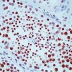
- Formalin-fixed, paraffin-embedded human testis stained with XRCC1 antibody using peroxidase-conjugate and AEC chromogen. Note nuclear staining of spermatocytes.

Supportive validation
- Submitted by
- Invitrogen Antibodies (provider)
- Main image

- Experimental details
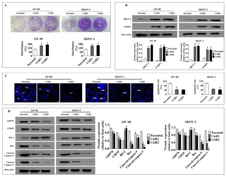
- Figure 5 The CisR OC activated DNA repair pathways and suppressed endoplasmic reticulum (ER)-stress mediated cell death. ( A ) The clonogenic growth rate analysis reveals a significantly faster clonogenic growth of CisR cells compared to parental cells. ( B ) Western blot analysis of DNA repair proteins, XRCC1 and PARP1, demonstrating significantly higher expression of DNA repair proteins in CisR cells compared to parental cells. ( C ) Apoptosis rate evaluated by Hoechst33342 staining shows significantly lower apoptosis in CisR cells compared to parental cells (Magnification, 40x, scale bar 20 um) ( D ) the Western blot analysis for GRP78, CHOP, Bcl-2, Bax, cleaved caspase-9 and cleaved caspase-3 shows significantly decreased levels of pro-apoptotic proteins and significantly increased levels of anti-apoptotic protein in CisR compared to parental cells. Values were represented as mean +- SD. * p < 0.05, # p < 0.01, compared with the parental group.
- Submitted by
- Invitrogen Antibodies (provider)
- Main image
- Experimental details
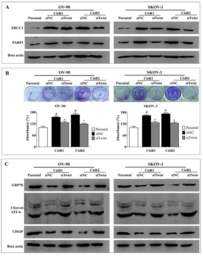
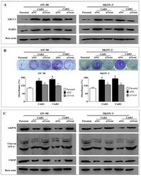
- Figure 8 Twist knockdown reduces cell growth potential in CisR OC cells. ( A ) Western blot analysis for DNA repair proteins, PARP1 and XRCC1. ( B ) The cell survival analysis assayed by clonogenic assay. ( C ) Western blot analysis for expression of ER stress signaling proteins, GRP78, cleaved ATF-6 and CHOP. Parental: non-transfected parental cells; siNC: cisplatin resistance cells transfected with non-targeting negative control siRNA; siTwist: cisplatin resistance cells transfected with Twist siRNA. Values were represented as mean +- SD. # p < 0.05, compared with the parental group and * p < 0.05, compared with siNC group.