Explore
 Validate
 Learn
Learn Western blot
Antibody data
- Antibody Data
- Antigen structure
- References [0]
- Comments [0]
- Validations
- Western blot [3]
- Immunohistochemistry [1]
Submit
Validation data
Reference
Comment
Report error
- Product number
- PA3-027 - Provider product page

- Provider
- Invitrogen Antibodies
- Product name
- GPR39 Polyclonal Antibody
- Antibody type
- Polyclonal
- Antigen
- Other
- Description
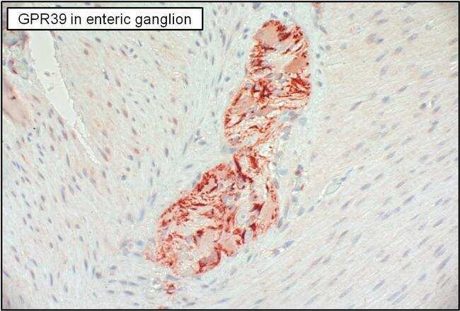
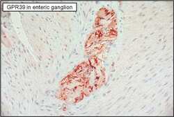
- IHC (P) analysis shows positive staining of GPR39 in human enteric ganglion.
- Concentration
- Conc. Not Determined
No comments: Submit comment
Supportive validation
- Submitted by
- Invitrogen Antibodies (provider)
- Main image

- Experimental details
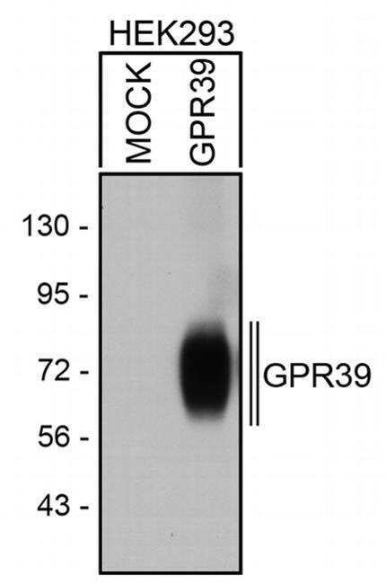
- Western blot analysis of GPR39 was performed by loading equal amounts of wheat germ lectin agarose bead enriched GPR receptor fractions from mock-transfected or GPR39 transfected HEK293 lysates onto a 7.5% Tris-HCl polyacrylamide gel. Proteins were transferred to a PVDF membrane, blocked and probed with a GPR39 polyclonal antibody (Product # PA3-027) at a dilution of 1:5000, overnight at 4C on a rocking platform, followed by an HRP-conjugated goat anti-rabbit IgG secondary antibody. Denatured GPR39 was detected at ~72kDa. Chemiluminescent detection was performed using ECL.
- Submitted by
- Invitrogen Antibodies (provider)
- Main image
- Experimental details
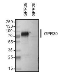
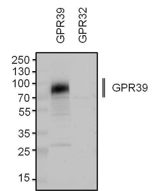
- Western blot analysis of GPR39 was performed by loading equal amounts of wheat germ lectin agarose bead enriched GPR receptor fractions from GPR39 or GPR25 transfected HEK293 lysates and 10 µL of PageRuler Plus Prestained Protein Ladder (Product # 26619) onto a 4-20% Tris-HCl polyacrylamide gel. Proteins were transferred to a PVDF membrane using the G2 Fast Blotter (Product # 62288), and blocked with StartingBlock T20 (TBS) Blocking Buffer (Product # 37543) for 1 hour at room temperature. GPR39 was detected at ~72 kDa using a GPR39 polyclonal antibody (Product # PA3-027) at a dilution of 1:2000 in StartingBlock T20 (TBS) Blocking Buffer (Product # 37543) overnight at 4C on a rocking platform, followed by an HRP-conjugated goat anti-rabbit IgG secondary antibody (Product # 31460) at a dilution of 1:20,000 for 1 hour. Chemiluminescent detection was performed using SuperSignal West Dura (Product # 34075). Images were acquired on a Thermo Scientific myECL Imager (Product # 62236).
- Submitted by
- Invitrogen Antibodies (provider)
- Main image
- Experimental details
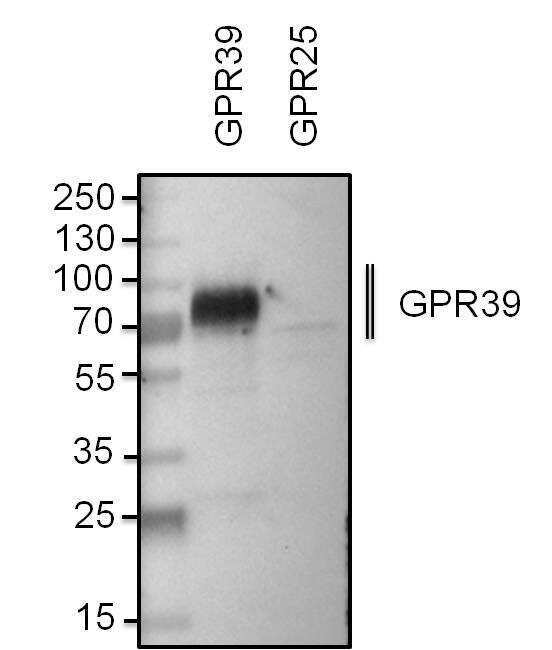
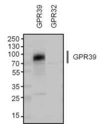
- Western blot analysis of GPR39 was performed by loading equal amounts of wheat germ lectin agarose bead enriched GPR receptor fractions from GPR39 or GPR32 transfected HEK293 lysates and 10 µL of PageRuler Plus Prestained Protein Ladder (Product # 26619) onto a 4-20% Tris-HCl polyacrylamide gel. Proteins were transferred to a PVDF membrane using the G2 Fast Blotter (Product # 62288), and blocked with StartingBlock T20 (TBS) Blocking Buffer (Product # 37543) for 1 hour at room temperature. GPR39 was detected at ~72 kDa using a GPR39 polyclonal antibody (Product # PA3-027) at a dilution of 1:1000 in StartingBlock T20 (TBS) Blocking Buffer (Product # 37543) overnight at 4C on a rocking platform, followed by an HRP-conjugated goat anti-rabbit IgG secondary antibody (Product # 31460) at a dilution of 1:20,000 for 1 hour. Chemiluminescent detection was performed using SuperSignal West Dura (Product # 34075). Images were acquired on a Thermo Scientific myECL Imager (Product # 62236).
Supportive validation
- Submitted by
- Invitrogen Antibodies (provider)
- Main image
- Experimental details
- Immunohistochemistry analysis of GPR39 was performed on human enteric ganglion tissue. To expose target proteins, antigen retrieval was performed by microwaving tissues for 20 minutes in 10mM sodium citrate buffer (pH 6.0). Tissue slides were probed with a GPR39 polyclonal antibody (Product # PA3-027) at a dilution of 1:3000, overnight at 4C in a humidified chamber. Tissues were washed, and detection was performed using an ABC kit composed of biotinylated goat anti-rabbit IgG, peroxidase-conjugated avidin, and 3-amino-9-ethylcarbazole (AEC) substrate in acetate buffer. Tissues were counterstained with hematoxylin and dehydrated to prep for mounting.