Explore
 Validate
 Learn
Learn Western blot
 Immunocytochemistry
Antibody data
- Antibody Data
- Antigen structure
- References [1]
- Comments [0]
- Validations
- Immunocytochemistry [3]
- Immunohistochemistry [3]
- Other assay [3]
Submit
Validation data
Reference
Comment
Report error
- Product number
- PA5-21976 - Provider product page

- Provider
- Invitrogen Antibodies
- Product name
- RPA70 Polyclonal Antibody
- Antibody type
- Polyclonal
- Antigen
- Recombinant full-length protein
- Description
- Recommended positive controls: 293T, A431, HeLa, HepG2, Mouse liver. Predicted reactivity: Mouse (87%), Rat (85%), Sheep (93%), Bovine (93%). Store product as a concentrated solution. Centrifuge briefly prior to opening the vial.
- Reactivity
- Human, Mouse
- Host
- Rabbit
- Isotype
- IgG
- Vial size
- 100 μL
- Concentration
- 0.4 mg/mL
- Storage
- Store at 4°C short term. For long term storage, store at -20°C, avoiding freeze/thaw cycles.
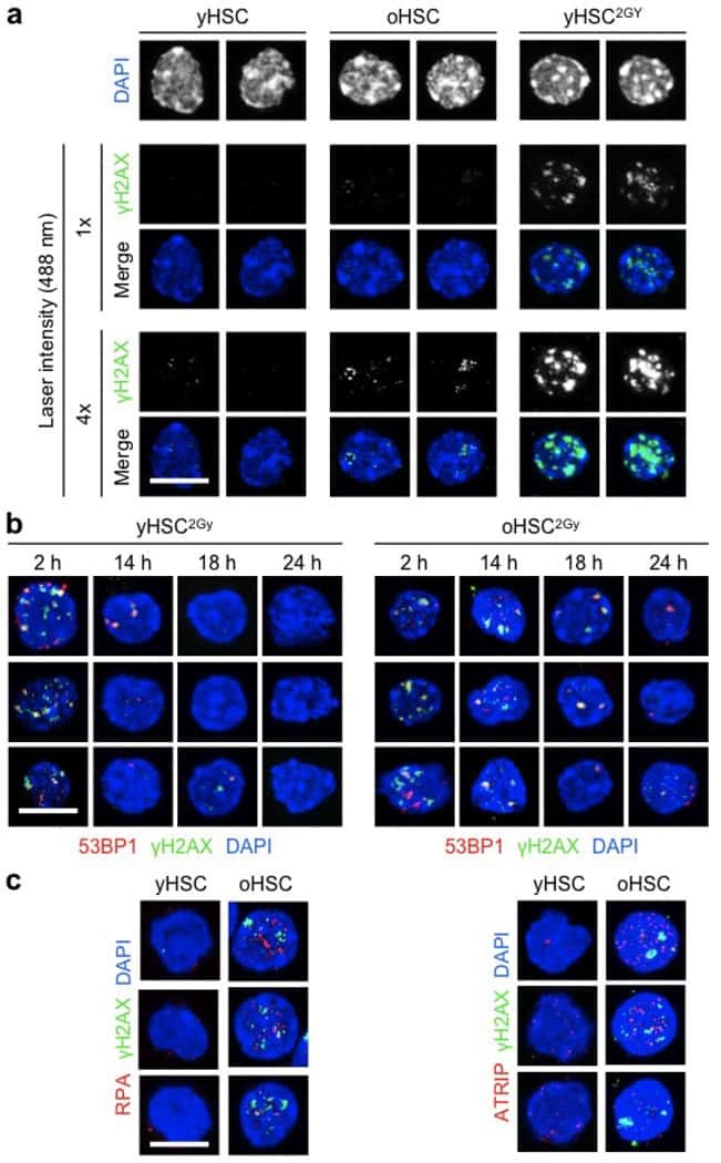
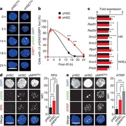
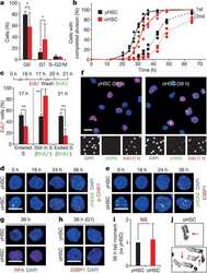
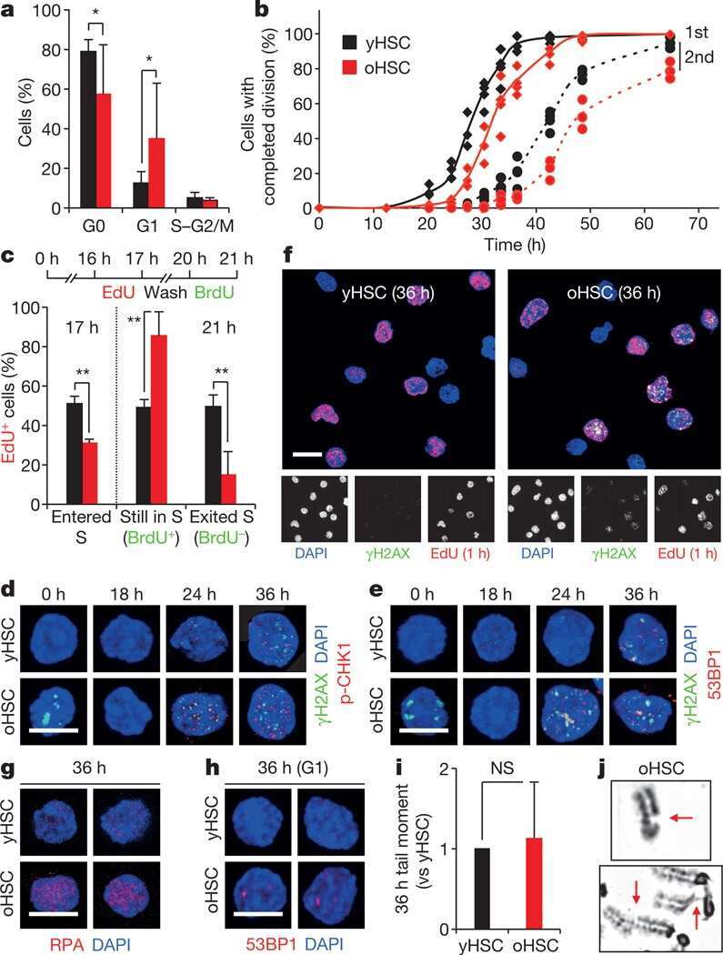
Submitted references Replication stress is a potent driver of functional decline in ageing haematopoietic stem cells.
Flach J, Bakker ST, Mohrin M, Conroy PC, Pietras EM, Reynaud D, Alvarez S, Diolaiti ME, Ugarte F, Forsberg EC, Le Beau MM, Stohr BA, Méndez J, Morrison CG, Passegué E
Nature 2014 Aug 14;512(7513):198-202
Nature 2014 Aug 14;512(7513):198-202
No comments: Submit comment
Supportive validation
- Submitted by
- Invitrogen Antibodies (provider)
- Main image
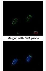
- Experimental details
- Immunofluorescent analysis of RPA 70 kDa subunit in paraformaldehyde-fixed HeLa cells using a RPA 70 kDa subunit polyclonal antibody (Product # PA5-21976) at a 1:200 dilution.

- Submitted by
- Invitrogen Antibodies (provider)
- Main image
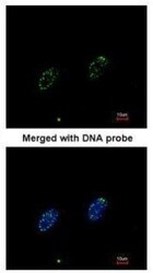
- Experimental details
- Immunofluorescence analysis of paraformaldehyde-fixed HeLa, using RPA 70 kDa subunit antibody (Product # PA5-21976) at 1:200 dilution.

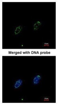
- Submitted by
- Invitrogen Antibodies (provider)
- Main image

- Experimental details
- Immunofluorescence analysis of paraformaldehyde-fixed HeLa, using RPA 70 kDa subunit antibody (Product # PA5-21976) at 1:200 dilution.
Supportive validation
- Submitted by
- Invitrogen Antibodies (provider)
- Main image
- Experimental details
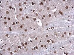
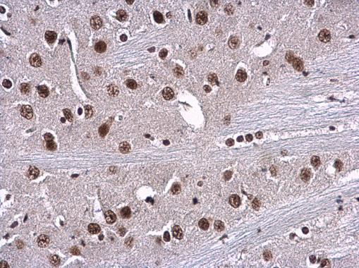
- RPA70 Polyclonal Antibody detects RPA1 protein at nucleus in mouse brain by immunohistochemical analysis. Sample: Paraffin-embedded mouse brain. RPA70 Polyclonal Antibody (Product # PA5-21976) diluted at 1:500. Antigen Retrieval: Citrate buffer, pH 6.0, 15 min.
- Submitted by
- Invitrogen Antibodies (provider)
- Main image
- Experimental details
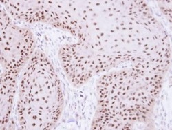
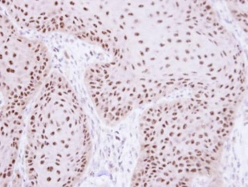
- Immunohistochemistry (Paraffin) analysis of RPA70 was performed in paraffin-embedded human lung papillary adenocarcinoma tissue using RPA70 Polyclonal Antibody (Product # PA5-21976) at a dilution of 1:250.

- Submitted by
- Invitrogen Antibodies (provider)
- Main image
- Experimental details
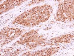
- Immunohistochemical analysis of paraffin-embedded Cal27 xenograft, using RPA 70 kDa subunit (Product # PA5-21976) antibody at 1:100 dilution. Antigen Retrieval: EDTA based buffer, pH 8.0, 15 min.
Supportive validation
- Submitted by
- Invitrogen Antibodies (provider)
- Main image
- Experimental details
- NULL

- Submitted by
- Invitrogen Antibodies (provider)
- Main image
- Experimental details
- NULL
- Submitted by
- Invitrogen Antibodies (provider)
- Main image
- Experimental details
- NULL