 Explore
 Validate
 Learn
Learn Western blot
 Immunohistochemistry
Antibody data
- Antibody Data
- Antigen structure
- References [2]
- Comments [0]
- Validations
- Western blot [1]
- Immunohistochemistry [1]
Submit
Validation data
Reference
Comment
Report error
- Product number
- HPA026744 - Provider product page

- Provider
- Atlas Antibodies
- Proper citation
- Atlas Antibodies Cat#HPA026744, RRID:AB_2194642
- Product name
- Anti-SPACA1
- Antibody type
- Polyclonal
- Description
- Polyclonal Antibody against Human SPACA1, Gene description: sperm acrosome associated 1, Alternative Gene Names: SAMP32, Validated applications: IHC, WB, Uniprot ID: Q9HBV2, Storage: Store at +4°C for short term storage. Long time storage is recommended at -20°C.
- Reactivity
- Human
- Host
- Rabbit
- Conjugate
- Unconjugated
- Isotype
- IgG
- Vial size
- 100 µl
- Concentration
- 0.1 mg/ml
- Storage
- Store at +4°C for short term storage. Long time storage is recommended at -20°C.
- Handling
- The antibody solution should be gently mixed before use.
Submitted references Case report of apoptosis in testis of four AZFc-deleted patients: increased DNA fragmentation during meiosis, but decreased apoptotic markers in post-meiotic germ cells
Complete deletion of the AZFb interval from the Y chromosome in an oligozoospermic man
Streichemberger E, Perrin J, Saias-Magnan J, Karsenty G, Malzac P, Grillo J, Mitchell M, Metzler-Guillemain C
Human Reproduction 2012;27(7):1939-1945
Human Reproduction 2012;27(7):1939-1945
Complete deletion of the AZFb interval from the Y chromosome in an oligozoospermic man
Longepied G, Saut N, Aknin-Seifer I, Levy R, Frances A, Metzler-Guillemain C, Guichaoua M, Mitchell M
Human Reproduction 2010;25(10):2655-2663
Human Reproduction 2010;25(10):2655-2663
No comments: Submit comment
Enhanced validation
- Submitted by
- Atlas Antibodies (provider)
- Enhanced method
- Recombinant expression validation
- Main image

- Experimental details
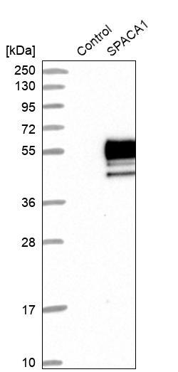
- Western blot analysis in control (vector only transfected HEK293T lysate) and SPACA1 over-expression lysate (Co-expressed with a C-terminal myc-DDK tag (~3.1 kDa) in mammalian HEK293T cells, LY410624).
- Sample type
- Human
- Protocol
- Protocol
Supportive validation
- Submitted by
- Atlas Antibodies (provider)
- Enhanced method
- Orthogonal validation
- Main image
- Experimental details
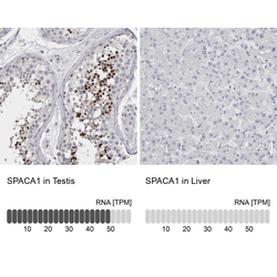
- Immunohistochemistry analysis in human testis and liver tissues using HPA026744 antibody. Corresponding SPACA1 RNA-seq data are presented for the same tissues.
- Sample type
- Human
- Protocol
- Protocol
