Explore
 Validate
 Learn
LearnPA5-111868
antibody from Invitrogen Antibodies
Targeting: GFRA1
GDNFR, GDNFRA, GFR-ALPHA-1, RET1L, RETL1, TRNR1
 Western blot
 Immunoelectron microscopy
Antibody data
- Antibody Data
- Antigen structure
- References [0]
- Comments [0]
- Validations
- Western blot [1]
- Immunocytochemistry [1]
- Immunohistochemistry [1]
Submit
Validation data
Reference
Comment
Report error
- Product number
- PA5-111868 - Provider product page

- Provider
- Invitrogen Antibodies
- Product name
- GFR alpha-1 (extracellular) Polyclonal Antibody
- Antibody type
- Polyclonal
- Antigen
- Synthetic peptide
- Description
- Excitation: 405 nm; Emission: 506 nm; Laser: Violet Laser.
- Reactivity
- Human, Mouse, Rat
- Host
- Rabbit
- Isotype
- IgG
- Vial size
- 50 µL
- Concentration
- 0.8 mg/mL
- Storage
- -20°C
No comments: Submit comment
Supportive validation
- Submitted by
- Invitrogen Antibodies (provider)
- Main image
- Experimental details
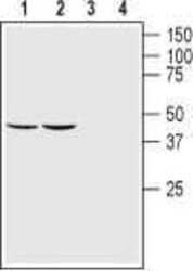
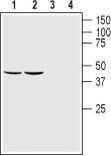
- Western Blot analysis of GFR alpha-1 was performed in rat (lanes 1 and 3) and mouse (lanes 2 and 4) brain lysates. Lane 1,2: GFR alpha-1 (extracellular) Antibody (Product # PA5-111868) at a dilution of 1:200. Lane 3,4: GFR alpha-1 (extracellular) Antibody, preincubated with the control peptide antigen.
Supportive validation
- Submitted by
- Invitrogen Antibodies (provider)
- Main image
- Experimental details
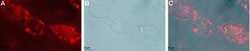
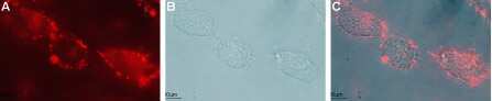
- Immunocytochemistry-Immunofluorescence analysis of GFR alpha-1 in live intact live intact rat C6 glioma cells using GFR alpha-1 (extracellular) Antibody (Product # PA5-111868) (1:50), followed by goat-anti-rabbit-DyLight-594 secondary antibody (red) (A). B) Live view of the cells. C) Merge of the two images.
Supportive validation
- Submitted by
- Invitrogen Antibodies (provider)
- Main image
- Experimental details
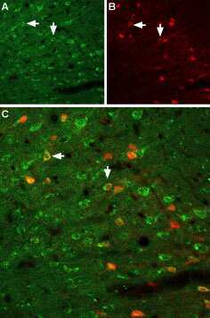
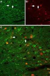
- Immunohistochemistry analysis of GFR alpha-1 in immersion-fixed, frozen rat neocortex tissue sections using GFR alpha-1 (extracellular) Antibody (Product # PA5-111868) at a dilution of 1:100. A) GFRA1 (green) is visualized in neocortex neurons. B) Neurons expressing y-amino butyric acid (GABA) are labeled with parvalbumin (red). C) Merge of the two images demonstrates partial colocalization (arrows).