 Explore
 Validate
 Learn
LearnANT-021-200UL
antibody from Invitrogen Antibodies
Targeting: GFRA1
GDNFR, GDNFRA, GFR-ALPHA-1, RET1L, RETL1, TRNR1
 Western blot
Antibody data
- Antibody Data
- Antigen structure
- References [0]
- Comments [0]
- Validations
- Western blot [2]
- Immunocytochemistry [2]
- Immunohistochemistry [1]
Submit
Validation data
Reference
Comment
Report error
- Product number
- ANT-021-200UL - Provider product page

- Provider
- Invitrogen Antibodies
- Product name
- GFR alpha 1 (extracellular) Polyclonal Antibody
- Antibody type
- Polyclonal
- Antigen
- Other
- Reactivity
- Human, Mouse, Rat
- Host
- Rabbit
- Isotype
- IgG
- Vial size
- 200 µL
- Concentration
- 0.8 mg/mL
- Storage
- -20° C, Avoid Freeze/Thaw Cycles
No comments: Submit comment
Supportive validation
- Submitted by
- Invitrogen Antibodies (provider)
- Main image
- Experimental details
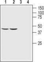
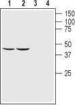
- Western blot analysis of rat (lanes 1 and 3) and mouse (lanes 2 and 4) brain lysates: - 1,2. Anti-GFR alpha 1 (extracellular) Antibody (#ANT-021), (1:200).3,4. Anti-GFR alpha 1 (extracellular) Antibody , preincubated with GFR alpha 1 (extracellular) Blocking Peptide (#BLP-NT021).

- Submitted by
- Invitrogen Antibodies (provider)
- Main image
- Experimental details
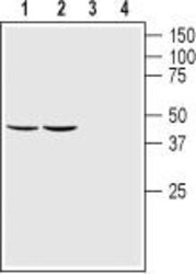
- Western blot analysis of rat (lanes 1 and 3) and mouse (lanes 2 and 4) brain lysates: - 1,2. Anti-GFR alpha 1 (extracellular) Antibody (#ANT-021), (1:200).3,4. Anti-GFR alpha 1 (extracellular) Antibody , preincubated with GFR alpha 1 (extracellular) Blocking Peptide (#BLP-NT021).
Supportive validation
- Submitted by
- Invitrogen Antibodies (provider)
- Main image
- Experimental details
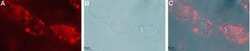
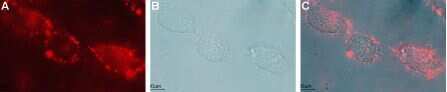
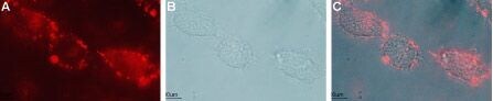
- Expression of GFRA1 in live intact rat C6 glioma cells - Cell surface detection of GFRA1 in live intact rat C6 glioma cells with Anti-GFR alpha 1 (extracellular) Antibody (#ANT-021), (1:50), followed by goat- Anti-rabbit-DyLight-594 secondary Antibody (red) (A). B. Live view of the cells. C. Merge of the two images.
- Submitted by
- Invitrogen Antibodies (provider)
- Main image

- Experimental details
- Expression of GFRA1 in live intact rat C6 glioma cells - Cell surface detection of GFRA1 in live intact rat C6 glioma cells with Anti-GFR alpha 1 (extracellular) Antibody (#ANT-021), (1:50), followed by goat- Anti-rabbit-DyLight-594 secondary Antibody (red) (A). B. Live view of the cells. C. Merge of the two images.
Supportive validation
- Submitted by
- Invitrogen Antibodies (provider)
- Main image
- Experimental details
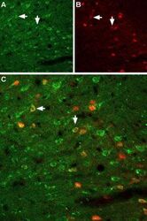
- Expression ofGFRA1 in rat neocortex - Immunohistochemical staining of immersion-fixed, free floating rat brain frozen sections using Anti-GFR alpha 1 (extracellular) Antibody (#ANT-021), (1:100). A. GFRA1 (green) is visualized in neocortex neurons. B. Neurons expressing gamma -amino butyric acid (GABA) are labeled with parvalbumin (red). C. Merge of the two images demonstrates partial colocalization (arrows).
