 Explore
 Validate
 Learn
LearnPA5-19873
antibody from Invitrogen Antibodies
Targeting: GFRA1
GDNFR, GDNFRA, GFR-ALPHA-1, RET1L, RETL1, TRNR1
 Western blot
Antibody data
- Antibody Data
- Antigen structure
- References [0]
- Comments [0]
- Validations
- Western blot [3]
- Immunocytochemistry [1]
- Immunohistochemistry [4]
Submit
Validation data
Reference
Comment
Report error
- Product number
- PA5-19873 - Provider product page

- Provider
- Invitrogen Antibodies
- Product name
- GFR alpha-1 Polyclonal Antibody
- Antibody type
- Polyclonal
- Antigen
- Synthetic peptide
- Description
- A suggested positive control is human brain tissue lysate.
- Concentration
- 1 mg/mL
No comments: Submit comment
Supportive validation
- Submitted by
- Invitrogen Antibodies (provider)
- Main image

- Experimental details
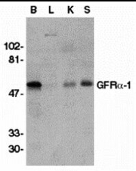
- Western blot analysis of crude membrane fractions of (B), liver (L), kidney (K), and spleen (S), respectively, using a GFR alpha 1 polyclonal antibody (Product # PA5-19873) at 1:500 dilution.

- Submitted by
- Invitrogen Antibodies (provider)
- Main image

- Experimental details
- Western Blot analysis of GFR alpha 1 in crude membrane fractions of human brain (B), liver (L), kidney (K), and spleen (S), respectively, with GFR alpha-1 Polyclonal Antibody (Product # PA5-19873) at 1 µg/mL.

- Submitted by
- Invitrogen Antibodies (provider)
- Main image
- Experimental details
- Western Blot analysis of GFR alpha 1 in crude membrane fractions of human brain (B), liver (L), kidney (K), and spleen (S), respectively, with GFR alpha-1 Polyclonal Antibody (Product # PA5-19873) at 1 µg/mL.

Supportive validation
- Submitted by
- Invitrogen Antibodies (provider)
- Main image
- Experimental details
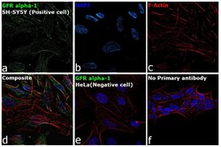
- Immunofluorescence analysis of GFR alpha-1 was performed using SH-SY5Y and HeLa cells. The cells were fixed with 4% paraformaldehyde for 10 minutes, permeabilized with 0.1% Triton™ X-100 for 15 minutes, and blocked with 2% BSA for 1 hour at room temperature. The cells were labeled with GFR alpha-1 Polyclonal Antibody (Product # PA5-19873) at 1:100 dilution in 0.1% BSA and incubated overnight at 4 degree and then labeled with Goat anti-Rabbit IgG (H+L) Highly Cross-Adsorbed Secondary Antibody, Alexa Fluor Plus 488 (Product # A32731) at a dilution of 1:2000 for 45 minutes at room temperature (Panel a: green). Nuclei (Panel b: blue) were stained with ProLong™ Diamond Antifade Mountant with DAPI (Product # P36962). F-actin (Panel c: red) was stained with Rhodamine Phalloidin (Product # R415, 1:300). Panel d represents the composite image showing cytoplasmic and membrane localization of GFR alpha-1. Panel e represents HeLa cells having no significant expression of GFR alpha 1. Panel f represents control SH-SY5Y cells with no primary antibody to assess background. The images were captured at 60X magnification.

Supportive validation
- Submitted by
- Invitrogen Antibodies (provider)
- Main image

- Experimental details
- Immunofluorescence of GFR alpha 1 in human brain tissue with GFR alpha-1 Polyclonal Antibody (Product # PA5-19873) at 20 µg/mL. Red: GFR alpha 1 Blue: DAPI staining

- Submitted by
- Invitrogen Antibodies (provider)
- Main image

- Experimental details
- Immunofluorescence of GFR alpha 1 in human brain tissue with GFR alpha-1 Polyclonal Antibody (Product # PA5-19873) at 20 µg/mL. Red: GFR alpha 1 Blue: DAPI staining

- Submitted by
- Invitrogen Antibodies (provider)
- Main image

- Experimental details
- Immunohistochemistry of GFR alpha 1 in human brain tissue with GFR alpha-1 Polyclonal Antibody (Product # PA5-19873) at 1 µg/mL.

- Submitted by
- Invitrogen Antibodies (provider)
- Main image

- Experimental details
- Immunohistochemistry of GFR alpha 1 in human brain tissue with GFR alpha-1 Polyclonal Antibody (Product # PA5-19873) at 2.5 µg/mL.
