Explore
 Validate
 Learn
Learn701395
antibody from Invitrogen Antibodies
Targeting: GZMB
CCPI, CGL-1, CGL1, CSP-B, CSPB, CTLA1, CTSGL1, HLP, SECT
 Western blot
Antibody data
- Antibody Data
- Antigen structure
- References [0]
- Comments [0]
- Validations
- Western blot [3]
- Immunocytochemistry [1]
- Flow cytometry [1]
Submit
Validation data
Reference
Comment
Report error
- Product number
- 701395 - Provider product page

- Provider
- Invitrogen Antibodies
- Product name
- Granzyme B Recombinant Rabbit Monoclonal Antibody (23H8L20)
- Antibody type
- Monoclonal
- Antigen
- Synthetic peptide
- Description
- Intact IgG appears on a non-reducing gel as ~150 kDa band and upon reduction generating a ~25 kDa light chain band and a ~50 kDa heavy chain.
- Antibody clone number
- 23H8L20
- Concentration
- 0.5 mg/mL
No comments: Submit comment
Supportive validation
- Submitted by
- Invitrogen Antibodies (provider)
- Main image

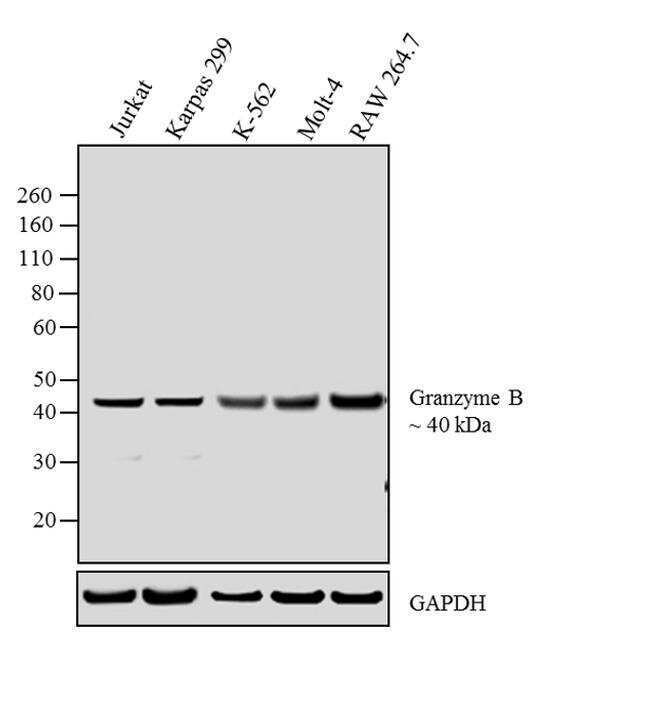
- Experimental details
- Western blot analysis was performed on whole cell extracts (30 µg lysate) of Jurkat (Lane 1), Karpas 299 (Lane 2), K-562 (Lane 3), Molt-4 (Lane 4) and RAW 264.7 (Lane 5).The blots were probed with Granzyme B Rabbit monoclonal Antibody (Product # 701395, 2 µg/mL) and detected by chemiluminescence using Goat anti-Rabbit IgG (H+L) Superclonal™ Secondary Antibody, HRP conjugate (Product # A27036, 0.4 µg/mL, 1:2500 dilution). A 40 kDa band corresponding to glycosylated form of Granzyme B was observed across the cell lines tested. Known quantity of protein samples were electrophoresed using Novex® NuPAGE® 4-12 % Bis-Tris gel (Product # NP0322BOX), XCell SureLock™ Electrophoresis System (Product # EI0002) and Novex® Sharp Pre-Stained Protein Standard (Product # LC5800). Resolved proteins were then transferred onto a nitrocellulose membrane with iBlot® 2 Dry Blotting System (Product # IB21001). The membrane was probed with the relevant primary and secondary Antibody following blocking with 5% skimmed milk. Chemiluminescent detection was performed using Pierce™ ECL Western Blotting Substrate (Product # 32106).
- Submitted by
- Invitrogen Antibodies (provider)
- Main image
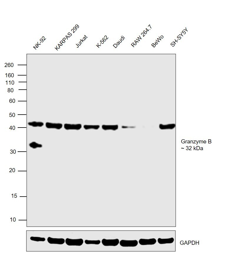
- Experimental details
- Western blot was performed using Anti-Granzyme B Recombinant Rabbit Monoclonal Antibody (23H8L20) (Product # 701395) and a 32 kDa band corresponding to Granzyme B was observed only in NK-92 cells which is reported to be positive and not in any other cell lines. Whole cell extracts (30 µg lysate) of NK-92 (Lane 1), KARPAS 299 (Lane 2), Jurkat (Lane 3), K-562 (Lane 4), Daudi (Lane 5), RAW 264.7 (Lane 6), BeWo (Lane 7) and SH-SY5Y (Lane 8) were electrophoresed using NuPAGE™ 10% Bis-Tris Protein Gel (Product # NP0302BOX). Resolved proteins were then transferred onto a nitrocellulose membrane (Product # IB23001) by iBlot® 2 Dry Blotting System (Product # IB21001). The blot was probed with the primary antibody (2 µg/mL) and detected by chemiluminescence with Goat anti-Rabbit IgG (H+L) Superclonal™ Recombinant Secondary Antibody, HRP (Product # A27036,1:4000 dilution) using the iBright FL 1000 (Product # A32752). Chemiluminescent detection was performed using Novex® ECL Chemiluminescent Substrate Reagent Kit (Product # WP20005). Uncharacterized band of ~ 40 kDa was also observed across the cell lines tested.
- Submitted by
- Invitrogen Antibodies (provider)
- Main image
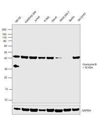
- Experimental details
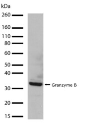
- Western blot analysis of Granzyme B in whole cell extracts from Jurkat using a Granzyme B recombinant rabbit monoclonal antibody (Product # 701395) at a dilution of 1 µg/mL. Detection was performed using an HRP-conjugated goat anti-rabbit secondary antibody followed by chemiluminescence (ECL). Results show a band at ~32kDa.

Supportive validation
- Submitted by
- Invitrogen Antibodies (provider)
- Main image
- Experimental details
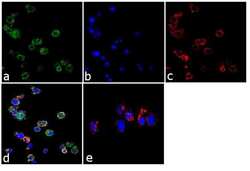
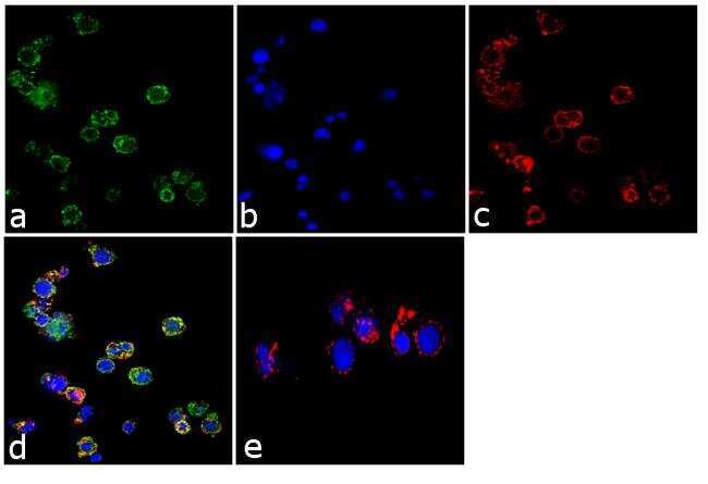
- Immunofluorescence analysis of Granzyme B was performed using 70% confluent log phase Jurkat cells. The cells were fixed with 4% paraformaldehyde for 10 minutes, permeabilized with 0.1% Triton™ X-100 for 10 minutes, and blocked with 1% BSA for 1 hour at room temperature. The cells were labeled with Granzyme B (23H8L20) Rabbit Monoclonal Antibody (Product # 701395) at 2 µg/mL in 0.1% BSA and incubated for 3 hours at room temperature and then labeled with Goat anti-Rabbit IgG (H+L) Superclonal™ Secondary Antibody, Alexa Fluor® 488 conjugate (Product # A27034) at a dilution of 1:2000 for 45 minutes at room temperature (Panel a: green). Nuclei (Panel b: blue) were stained with SlowFade® Gold Antifade Mountant with DAPI (Product # S36938). F-actin (Panel c: red) was stained with Alexa Fluor® 555 Rhodamine Phalloidin (Product # R415, 1:300). Panel d represents the merged image showing cytoplasmic localization. Panel e shows the control without primary antibody. The images were captured at 60X magnification.
Supportive validation
- Submitted by
- Invitrogen Antibodies (provider)
- Main image
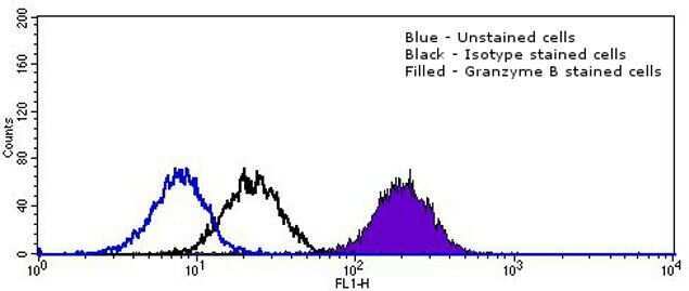
- Experimental details
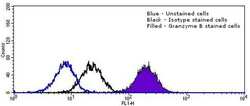
- Flow cytometry analysis of Granzyme B in Jurkat cells using a Granzyme B recombinant rabbit monoclonal antibody (Product # 701395). Cells were fixed and permeabilized using FIX & PERM (Product # GAS-004) reagent, and detection was performed using an Alexa Fluor 488 goat anti-rabbit IgG (right peak) compared to an isotype control (middle peak, black) and a control without primary antibody (left peak, blue).