Explore
 Validate
 Learn
Learn58-8896-42
antibody from Invitrogen Antibodies
Targeting: GZMB
CCPI, CGL-1, CGL1, CSP-B, CSPB, CTLA1, CTSGL1, HLP, SECT
 Flow cytometry
Antibody data
- Antibody Data
- Antigen structure
- References [1]
- Comments [0]
- Validations
- Flow cytometry [1]
- Other assay [2]
Submit
Validation data
Reference
Comment
Report error
- Product number
- 58-8896-42 - Provider product page

- Provider
- Invitrogen Antibodies
- Product name
- Granzyme B Monoclonal Antibody (N4TL33), Alexa Fluor™ 532, eBioscience™
- Antibody type
- Monoclonal
- Antigen
- Other
- Description
- Applications Reported: This N4TL33 antibody has been reported for use in intracellular staining followed by flow cytometric analysis. Applications Tested: This N4TL33 antibody has been pre-diluted and tested by intracellular staining followed by flow cytometric analysis of normal human peripheral blood cells. This may be used at 5 µL (0.5 µg) per test. A test is defined as the amount (µg) of antibody that will stain a cell sample in a final volume of 100 µL. Cell number should be determined empirically but can range from 10^5 to 10^8 cells/test. Alexa Fluor™ 532 is excited with the Green laser (532 nm) and emits at 561 nm. This cannot be used with the Yellow-Green laser (561 nm). We recommend using a 560/14 band pass filter. Please make sure that your instrument is capable of detecting this fluorochrome. Excitation: 532 nm; Emission: 561 nm; Laser: Green Laser
- Reactivity
- Human
- Host
- Mouse
- Conjugate
- Yellow dye
- Isotype
- IgG
- Antibody clone number
- N4TL33
- Vial size
- 100 Tests
- Concentration
- 5 µL/Test
- Storage
- 4° C, store in dark, DO NOT FREEZE!
Submitted references LINC01123 promotes immune escape by sponging miR-214-3p to regulate B7-H3 in head and neck squamous-cell carcinoma.
Li H, Yang Z, Yang X, Zhang F, Wang J, Wu Z, Wanyan C, Meng Q, Gao W, Yang X, Wei J
Cell death & disease 2022 Feb 3;13(2):109
Cell death & disease 2022 Feb 3;13(2):109
No comments: Submit comment
Supportive validation
- Submitted by
- Invitrogen Antibodies (provider)
- Main image

- Experimental details
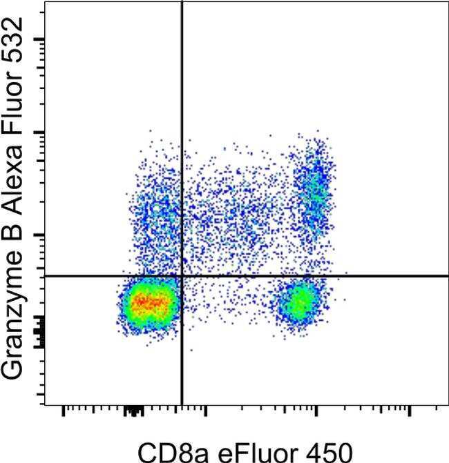
- Normal human peripheral blood cells were stained intracellularly, using the Intracellular Fixation & Permeabilization Buffer Set (Product # 88-8824-00) and protocol, with CD8a Monoclonal Antibody, eFluor 450 (Product # 48-0088-82) and Granzyme B Monoclonal Antibody, Alexa Fluor 532. Cells in the lymphocyte gate were used for analysis.
- Conjugate
- Yellow dye
Supportive validation
- Submitted by
- Invitrogen Antibodies (provider)
- Main image
- Experimental details
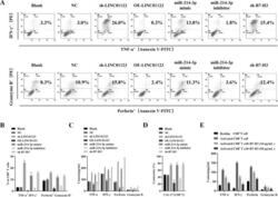
- Fig. 4 Upregulation of LINC01123 or B7-H3 or downregulation of miR-214-3p induces dysfunction of CD8 + T cells. CD8 + T cells were isolated from PBMCs using immunomagnetic beads. After activation and amplification, CD8 + T cells were cocultured with different CAL-27 cell treatment groups. A Expression of TNF-alpha, IFN-gamma, perforin, and granzyme B in CD8 + T-cell population was analyzed with flow cytometry. B Statistical analysis of the results from flow cytometry of CD8 + T cells. C Expression levels of TNF-alpha, IFN-gamma, perforin, and granzyme B in CAL-27-CD8 + T cocultured supernatants, as detected by ELISA. D CCK8 cytotoxicity test of CD8 + T cells. E ELISA was used to assess the immune activity of CD8 + T cells after recombinant human B7-H3 treatment. * P < 0.05 compared with cells without treatment. The experiment was repeated three times.
- Conjugate
- Yellow dye

- Submitted by
- Invitrogen Antibodies (provider)
- Main image
- Experimental details
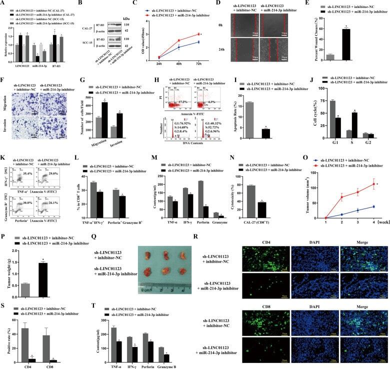
- Fig. 6 The function of LINC01123 in HNSCC can be reversed by miR-214-3p silencing. HNSCC cells were transfected with sh-LINC01123 in the presence of inhibitor-NC or miR-214-3p inhibitor. A Relative expression levels of LINC01123, miR-214-3p, and B7-H3 were determined by qRT-PCR. B B7-H3 protein expression was determined by western blot analysis. C CAL-27 cell viability was measured using a CCK8 assay. D Migration of CAL-27 cells was determined by scratch wound-healing assay. E Statistical results of the scratch-healing test. F Migration and invasion of CAL-27 cells, as determined by transwell migration and invasion assay. G Number of migration and invasion cells. H Apoptosis rate and cell-cycle stage were detected by flow cytometry. I Statistical analysis of apoptosis results. J Statistical analysis of cell cycle stage results. K CD8 + T cells were cocultured with CAL-27 cells that had been transfected with plasmids, and the expression of TNF-alpha, IFN-gamma, perforin, and granzyme B in the CD8 + T cells was analyzed by flow cytometry. L Statistical analysis of CD8 + T-cell flow cytometry results. M After different treatments, CAL-27 cells were cocultured with CD8 + T cells, and the expression levels of TNF-alpha, IFN-gamma, perforin, and granzyme B in coculture supernatants were detected by ELISA. N Killing effect of CD8 + T cells on CAL-27 cells, as assessed by CCK8. O Tumor-volume growth curves of the sh-LINC01123 + inhibitor-NC group and sh-LINC01123 + miR-214-3p-inhibit
- Conjugate
- Yellow dye