Explore
 Validate
 Learn
Learn Western blot
 ELISA
ELISAAntibody data
- Antibody Data
- Antigen structure
- References [1]
- Comments [0]
- Validations
- Western blot [1]
- Immunocytochemistry [4]
- Immunohistochemistry [4]
Submit
Validation data
Reference
Comment
Report error
- Product number
- PA5-97022 - Provider product page

- Provider
- Invitrogen Antibodies
- Product name
- MDC1 Polyclonal Antibody
- Antibody type
- Polyclonal
- Antigen
- Recombinant full-length protein
- Description
- Immunogen sequence: MEDTQAIDWD VEEEEETEQS SESLRCNVEP VGRLHIFSGA HGPEKDFPLH LGKNVVGRMP DCSVALPFPS ISKQHAEIEI LAWDKAPILR DCGSLNGTQI LRPPKVLSPG VSHRLRDQEL ILFADLLCQY HRLDVSLPFV SRGPLTVEET PRVQGETQPQ RLLLAEDSEE EVDFLSERRM VKKSRTTSSS VIVPESDEEG HSPVLGGLGP PFAFNLNSDT DVEEGQQPAT EEASSAARRG ATVEAKQSEA EVVTEIQLEK DQPLVKERDN DTKVKRGAGN GVVPAGVILE RSQPPGEDSD TDVDDDSRPP GRPAEVHLER AQPFGFIDSD TDAEEERIPA TPVVIPMKKR; Positive Samples: Jurkat, LO2; Cellular Location: Chromosome, Nucleus
- Reactivity
- Human
- Host
- Rabbit
- Isotype
- IgG
- Vial size
- 100 μL
- Concentration
- 0.28 mg/mL
- Storage
- -20°C, Avoid Freeze/Thaw Cycles
Submitted references O-GlcNAcylation Affects the Pathway Choice of DNA Double-Strand Break Repair.
Averbek S, Jakob B, Durante M, Averbeck NB
International journal of molecular sciences 2021 May 27;22(11)
International journal of molecular sciences 2021 May 27;22(11)
No comments: Submit comment
Supportive validation
- Submitted by
- Invitrogen Antibodies (provider)
- Main image
- Experimental details
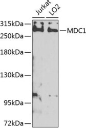
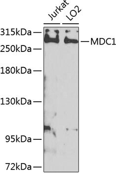
- Western blot analysis of MDC1 in extracts of various cell lines. Samples were incubated with MDC1 Polyclonal antibody (Product # PA5-97022) using a dilution of 1:1,000, followed by HRP Goat Anti-Rabbit IgG (H+L) at a dilution of 1:10,000. Lysates/proteins: 25 µg per lane. Blocking buffer: 3% nonfat dry milk in TBST. Detection: ECL Enhanced Kit. Exposure time: 5s.
Supportive validation
- Submitted by
- Invitrogen Antibodies (provider)
- Main image

- Experimental details
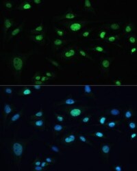
- Immunocytochemistry-Immunofluorescence analysis of MDC1 was performed in U-2 OS cells using MDC1 Polyclonal Antibody (Product # PA5-97022) at a dilution of 1:100. Blue: DAPI for nuclear staining.

- Submitted by
- Invitrogen Antibodies (provider)
- Main image

- Experimental details
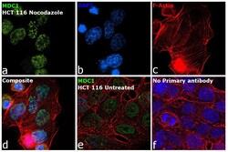
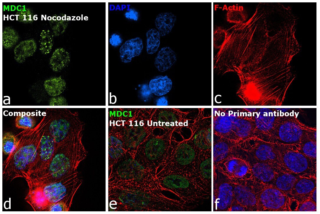
- Immunofluorescence analysis of Mediator of DNA damage checkpoint protein 1 was performed using 70% confluent log phase HCT 116 Nocodazole treated (200 nmol/L for 16hrs). The cells were fixed with 4% paraformaldehyde for 10 minutes, permeabilized with 0.1% Triton™ X-100 for 15 minutes, and blocked with 2% BSA for 45 minutes at room temperature. The cells were labeled with MDC1 Polyclonal Antibody (Product # PA5-97022) at 1:100 in 0.1% BSA, incubated at 4 degree celsius overnight, and then labeled with Donkey anti-Rabbit IgG (H+L) Highly Cross-Adsorbed Secondary Antibody, Alexa Fluor Plus 488 (Product # A32790), (1:2000 dilution), for 45 minutes at room temperature (Panel a: Green). Nuclei (Panel b: Blue) were stained with ProLong™ Diamond Antifade Mountant with DAPI (Product # P36962). F-actin (Panel c: Red) was stained with Rhodamine Phalloidin (Product # R415, 1:300). Panel d represents the merged image showing Nuclear foci localization. Panel e represents untreated cells with nuclear localization. Panel f represents control cells with no primary antibody to assess the background. The images were captured at 60X magnification.

- Submitted by
- Invitrogen Antibodies (provider)
- Main image
- Experimental details
- Immunofluorescence analysis of Mediator of DNA damage checkpoint protein 1 was performed using 70% confluent log phase HCT 116 Nocodazole treated (200 nmol/L for 16hrs). The cells were fixed with 4% paraformaldehyde for 10 minutes, permeabilized with 0.1% Triton™ X-100 for 15 minutes, and blocked with 2% BSA for 45 minutes at room temperature. The cells were labeled with MDC1 Polyclonal Antibody (Product # PA5-97022) at 1:100 in 0.1% BSA, incubated at 4 degree celsius overnight, and then labeled with Donkey anti-Rabbit IgG (H+L) Highly Cross-Adsorbed Secondary Antibody, Alexa Fluor Plus 488 (Product # A32790), (1:2000 dilution), for 45 minutes at room temperature (Panel a: Green). Nuclei (Panel b: Blue) were stained with ProLong™ Diamond Antifade Mountant with DAPI (Product # P36962). F-actin (Panel c: Red) was stained with Rhodamine Phalloidin (Product # R415, 1:300). Panel d represents the merged image showing Nuclear foci localization. Panel e represents untreated cells with nuclear localization. Panel f represents control cells with no primary antibody to assess the background. The images were captured at 60X magnification.
- Submitted by
- Invitrogen Antibodies (provider)
- Main image
- Experimental details
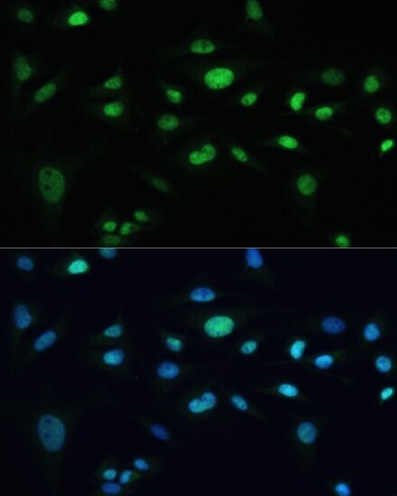
- Immunofluorescence analysis of MDC1 in U-2 OS cells. Samples were incubated with MDC1 Polyclonal antibody (Product # PA5-97022) using a dilution of 1:100. Blue: DAPI for nuclear staining.
Supportive validation
- Submitted by
- Invitrogen Antibodies (provider)
- Main image
- Experimental details
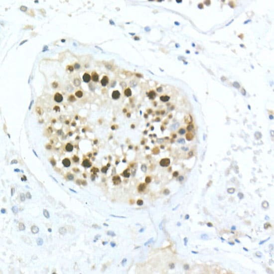
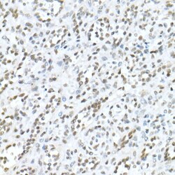
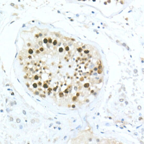
- Immunohistochemistry analysis of MDC1 in paraffin-embedded human spleen. Samples were incubated with MDC1 Polyclonal antibody (Product # PA5-97022) using a dilution of 1:20 (40x lens). Perform high pressure antigen retrieval with 10 mM citrate buffer pH 6.0 before commencing with IHC staining protocol.

- Submitted by
- Invitrogen Antibodies (provider)
- Main image

- Experimental details
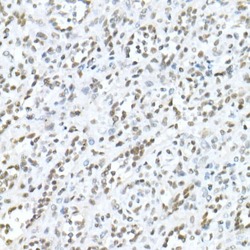
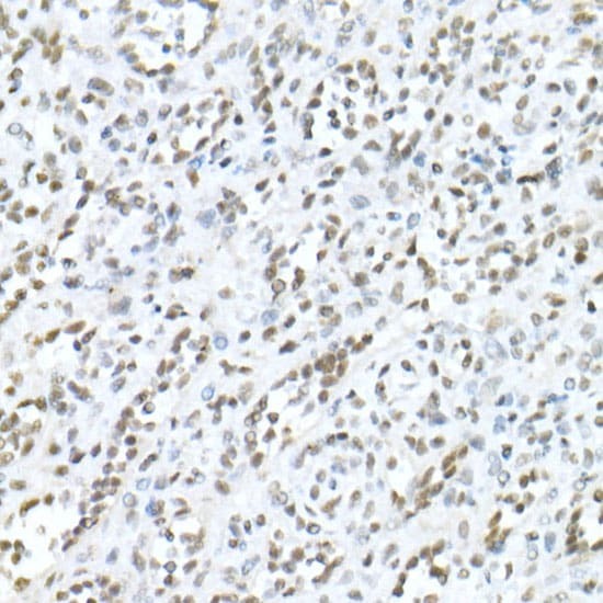
- Immunohistochemistry analysis of MDC1 in paraffin-embedded human testis. Samples were incubated with MDC1 Polyclonal antibody (Product # PA5-97022) using a dilution of 1:20 (40x lens). Perform high pressure antigen retrieval with 10 mM citrate buffer pH 6.0 before commencing with IHC staining protocol.
- Submitted by
- Invitrogen Antibodies (provider)
- Main image
- Experimental details
- Immunohistochemistry analysis of MDC1 in paraffin-embedded human spleen. Samples were incubated with MDC1 Polyclonal antibody (Product # PA5-97022) using a dilution of 1:20 (40x lens). Perform high pressure antigen retrieval with 10 mM citrate buffer pH 6.0 before commencing with IHC staining protocol.
- Submitted by
- Invitrogen Antibodies (provider)
- Main image

- Experimental details
- Immunohistochemistry analysis of MDC1 in paraffin-embedded human testis. Samples were incubated with MDC1 Polyclonal antibody (Product # PA5-97022) using a dilution of 1:20 (40x lens). Perform high pressure antigen retrieval with 10 mM citrate buffer pH 6.0 before commencing with IHC staining protocol.