 Explore
 Validate
 Learn
Learn Flow cytometry
Antibody data
- Antibody Data
- Antigen structure
- References [1]
- Comments [0]
- Validations
- Flow cytometry [2]
Submit
Validation data
Reference
Comment
Report error
- Product number
- FAB6198A-100UG - Provider product page

- Provider
- R&D Systems
- Product name
- Mouse CXCR5 APC-conjugated Antibody
- Antibody type
- Monoclonal
- Description
- Protein A or G purified from hybridoma culture supernatant. Detects recombinant mouse (rm) CXCR5 in direct ELISAs. In this format, no cross-reactivity with rmCXCR7 is observed.
- Reactivity
- Mouse
- Host
- Rat
- Conjugate
- Red dye
- Antigen sequence
Q04683- Isotype
- IgG
- Antibody clone number
- 614641
- Vial size
- 100 ug
- Storage
- Protect from light. Do not freeze. 12 months from date of receipt, 2 to 8 °C as supplied.
Submitted references Inhibition of increased circulating Tfh cell by anti-CD20 monoclonal antibody in patients with type 1 diabetes.
Xu X, Shi Y, Cai Y, Zhang Q, Yang F, Chen H, Gu Y, Zhang M, Yu L, Yang T
PloS one 2013;8(11):e79858
PloS one 2013;8(11):e79858
No comments: Submit comment
Supportive validation
- Submitted by
- R&D Systems (provider)
- Main image
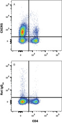
- Experimental details
- Detection of CXCR5 in Mouse T Follicular Helper Cells by Flow Cytometry. Mouse T follicular helper cells from immunized Balb/c mice were stained with Rat Anti-Mouse CD4 Alexa Fluor® 405-conjugated Monoclonal Antibody (Catalog # FAB554V) and either (A) Rat Anti-Mouse CXCR5 APC-conjugated Monoclonal Antibody (Catalog # FAB6198A) or (B) Rat IgG2A Allophycocyanin Isotype Control (Catalog # IC006A). View our protocol for Staining Membrane-associated Proteins.

- Submitted by
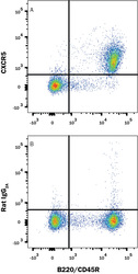
- R&D Systems (provider)
- Main image
- Experimental details
- Detection of CXCR5 in Mouse Splenocytes by Flow Cytometry. Mouse splenocytes were stained with Rat Anti-Mouse B220/CD45R PE-conjugated Monoclonal Antibody (Catalog # FAB1217P) and either (A) Rat Anti-Mouse CXCR5 APC-conjugated Monoclonal Antibody (Catalog # FAB6198A) or (B) Rat IgG2A Allophycocyanin Isotype Control (Catalog # IC006A). View our protocol for Staining Membrane-associated Proteins.
