Explore
 Validate
 Learn
Learn Western blot
 Immunohistochemistry
Antibody data
- Antibody Data
- Antigen structure
- References [0]
- Comments [0]
- Validations
- Immunohistochemistry [3]
- Flow cytometry [3]
Submit
Validation data
Reference
Comment
Report error
- Product number
- MA5-42534 - Provider product page

- Provider
- Invitrogen Antibodies
- Product name
- FEN1 Monoclonal Antibody (A1B12)
- Antibody type
- Monoclonal
- Antigen
- Recombinant full-length protein
- Description
- Positive Control: MCF-7 cell lysate, K562 cell lysate, Daudi cell lysate, PC-12 cell lysate, NIH/3T3 cell lysate, human tonsil tissue, human liver carcinoma tissue, human breast carcinoma tissue, K562. Subcellular Location: Nucleolus, nucleoplasm. Mitochondrion.
- Reactivity
- Human, Mouse, Rat
- Host
- Mouse
- Isotype
- IgG
- Antibody clone number
- A1B12
- Vial size
- 100 μL
- Concentration
- 2 mg/mL
- Storage
- Store at 4°C short term. For long term storage, store at -20°C, avoiding freeze/thaw cycles.
No comments: Submit comment
Supportive validation
- Submitted by
- Invitrogen Antibodies (provider)
- Main image
- Experimental details
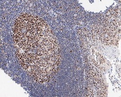
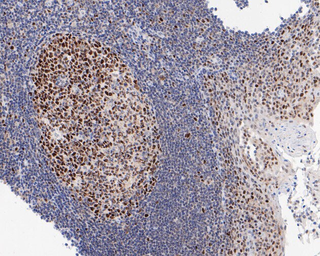
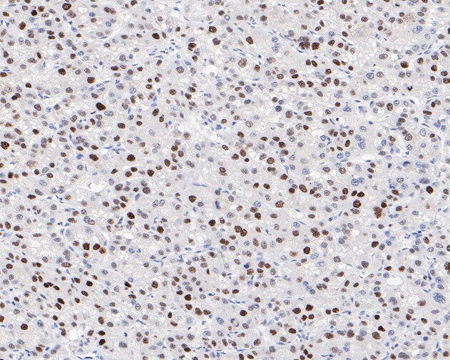
- Immunohistochemistry (Paraffin) analysis of paraffin-embedded human tonsil tissue using FEN1 Monoclonal Antibody (Product # MA5-42534). The section was pre-treated using heat mediated antigen retrieval with sodium citrate buffer (pH 6.0) for 20 minutes. The tissues were blocked in 5% BSA for 30 minutes at room temperature, washed with ddH2O and PBS, and then probed with the FEN1 antibody at a dilution of 1:400 for 30 minutes at room temperature. The detection was performed using an HRP conjugated compact polymer system. DAB was used as the chromogen. Tissues were counterstained with hematoxylin and mounted with DPX.
- Submitted by
- Invitrogen Antibodies (provider)
- Main image
- Experimental details
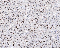
- Immunohistochemistry (Paraffin) analysis of paraffin-embedded human liver carcinoma tissue using FEN1 Monoclonal Antibody (Product # MA5-42534). The section was pre-treated using heat mediated antigen retrieval with sodium citrate buffer (pH 6.0) for 20 minutes. The tissues were blocked in 5% BSA for 30 minutes at room temperature, washed with ddH2O and PBS, and then probed with the FEN1 antibody at a dilution of 1:400 for 30 minutes at room temperature. The detection was performed using an HRP conjugated compact polymer system. DAB was used as the chromogen. Tissues were counterstained with hematoxylin and mounted with DPX.
- Submitted by
- Invitrogen Antibodies (provider)
- Main image
- Experimental details
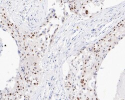
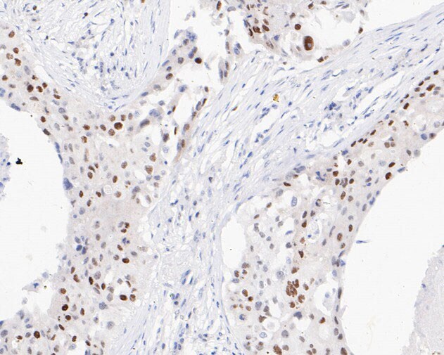
- Immunohistochemistry (Paraffin) analysis of paraffin-embedded human breast carcinoma tissue using FEN1 Monoclonal Antibody (Product # MA5-42534). The section was pre-treated using heat mediated antigen retrieval with sodium citrate buffer (pH 6.0) for 20 minutes. The tissues were blocked in 5% BSA for 30 minutes at room temperature, washed with ddH2O and PBS, and then probed with the FEN1 antibody at a dilution of 1:400 for 30 minutes at room temperature. The detection was performed using an HRP conjugated compact polymer system. DAB was used as the chromogen. Tissues were counterstained with hematoxylin and mounted with DPX.
Supportive validation
- Submitted by
- Invitrogen Antibodies (provider)
- Main image
- Experimental details
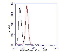
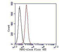
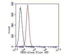
- Flow Cytometry analysis of FEN1 in K562 cells using FEN1 Monoclonal Antibody (Product # MA5-42534) at 1:50 (red). After incubation of the primary antibody at room temperature for an hour, the cells were stained with a Alexa Fluor 488-conjugated Goat anti-Mouse IgG Secondary antibody at 1:1,000 dilution for 30 minutes. Unlabelled sample was used as a control (cells without incubation with primary antibody; black).
- Submitted by
- Invitrogen Antibodies (provider)
- Main image
- Experimental details
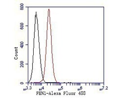
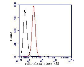
- Flow Cytometry analysis of FEN1 in K562 cells using FEN1 Monoclonal Antibody (Product # MA5-42534) at 1:50 (red). After incubation of the primary antibody at room temperature for an hour, the cells were stained with a Alexa Fluor 488-conjugated Goat anti-Mouse IgG Secondary antibody at 1:1,000 dilution for 30 minutes. Unlabelled sample was used as a control (cells without incubation with primary antibody; black).
- Submitted by
- Invitrogen Antibodies (provider)
- Main image
- Experimental details
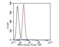
- Flow Cytometry analysis of FEN1 in K562 cells using FEN1 Monoclonal Antibody (Product # MA5-42534) at 1:50 (red). After incubation of the primary antibody at room temperature for an hour, the cells were stained with a Alexa Fluor 488-conjugated Goat anti-Mouse IgG Secondary antibody at 1:1,000 dilution for 30 minutes. Unlabelled sample was used as a control (cells without incubation with primary antibody; black).