Explore
 Validate
 Learn
Learn Western blot
Antibody data
- Antibody Data
- Antigen structure
- References [0]
- Comments [0]
- Validations
- Western blot [1]
- Immunocytochemistry [1]
Submit
Validation data
Reference
Comment
Report error
- Product number
- 711442 - Provider product page

- Provider
- Invitrogen Antibodies
- Product name
- FXR1 Recombinant Polyclonal Antibody
- Antibody type
- Polyclonal
- Antigen
- Other
- Reactivity
- Human, Mouse, Rat
- Host
- Rabbit
- Isotype
- IgG
- Vial size
- 100 µg
- Concentration
- 0.5 mg/mL
- Storage
- Store at 4°C short term. For long term storage, store at -20°C, avoiding freeze/thaw cycles.
No comments: Submit comment
Supportive validation
- Submitted by
- Invitrogen Antibodies (provider)
- Main image

- Experimental details
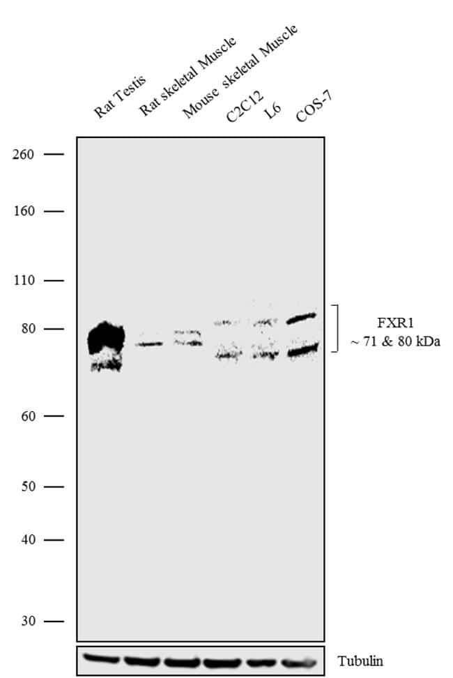
- Western blot analysis was performed on Tissue extracts (30 µg lysate) of Rat Testis (Lane 1), Rat Skeletal Muscle (Lane 2), Mouse Skeletal Muscle (Lane 3) and Whole cell extracts of C2C12 (Lane 4), L6 (Lane 5) and COS-7 (Lane 6). The blots were probed with Anti-FXR1 Recombinant Rabbit Polyclonal Antibody (Product # 711442, 1-2 µg/mL) and detected by chemiluminescence using Goat anti-Rabbit IgG (H+L) Superclonal Secondary Antibody, HRP conjugate (Product # A27036, 0.4 µg/mL, 1:2500 dilution). Two isoforms of 71 kDa and 80 kDa band corresponding to FXR1 was observed across the tissues and cell lines tested. Known quantity of protein samples were electrophoresed using Novex®NuPAGE®4-12% Bis-Tris gel (Product # NP0321BOX), XCell SureLock Electrophoresis System (Product # EI0002) and Novex® Sharp Pre-Stained Protein Standard (Product # LC5800). Resolved proteins were then transferred onto a nitrocellulose membrane with iBlot® Dry Blotting System (Product # IB21001). The membrane was probed with the relevant primary and secondary Antibody following blocking with 5% skimmed milk. Chemiluminescent detection was performed using Pierce™ ECL Western blotting Substrate (Product # 32106).
Supportive validation
- Submitted by
- Invitrogen Antibodies (provider)
- Main image

- Experimental details
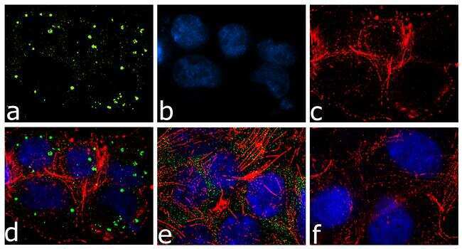
- For immunofluorescence analysis, A375 cells were fixed and permeabilized for detection of endogenous FXR1 using Anti-FXR1 Recombinant Rabbit Polyclonal Antibody (Product # 711442, 2 µg/mL) and labeled with Goat anti-Rabbit IgG (H+L) Superclonal Secondary Antibody, Alexa Fluor® 488 conjugate (Product # A27034, 1:2000). Panel a) shows representative cells that were stained for detection and localization of FXR1 protein (green), Panel b) is stained for nuclei (blue) using SlowFade® Gold Antifade Mountant with DAPI (Product # S36938). Panel c) represents cytoskeletal F-actin staining using Alexa Fluor® 555 Rhodamine Phalloidin (Product # R415, 1:300). Panel d) is a composite image of Panels a, b and c clearly demonstrating FXR1 localization in stress granules (green) in cells treated with MG132 (25 uM, 5 hrs). Panel e) represents untreated cells with diffused cytoplasmic staining. Panel f) represents control cells with no primary antibody to assess background. The images were captured at 60X magnification.