 Explore
 Validate
 Learn
Learn Western blot
 Immunoprecipitation
Antibody data
- Antibody Data
- Antigen structure
- References [0]
- Comments [0]
- Validations
- Western blot [2]
- Immunocytochemistry [1]
- Other assay [1]
Submit
Validation data
Reference
Comment
Report error
- Product number
- 702410 - Provider product page

- Provider
- Invitrogen Antibodies
- Product name
- FXR1 Recombinant Rabbit Monoclonal Antibody (6H25L10)
- Antibody type
- Monoclonal
- Antigen
- Other
- Reactivity
- Human, Mouse
- Host
- Rabbit
- Isotype
- IgG
- Antibody clone number
- 6H25L10
- Vial size
- 100 µg
- Concentration
- 0.5 mg/mL
- Storage
- Store at 4°C short term. For long term storage, store at -20°C, avoiding freeze/thaw cycles.
No comments: Submit comment
Supportive validation
- Submitted by
- Invitrogen Antibodies (provider)
- Main image

- Experimental details
- Knockdown of FXR1 was achieved by transfecting NIH3T3 cells with FXR1 specific siRNA (Silencer® select Cat # s15610 and s15612). Western blot analysis (Fig a) was performed using Whole cell extract from the FXR1 knock down cells (lane 3), non-specific scrambled siRNA transfected cells (lane 2) and untransfected cells (lane 1). The blots were probed with Anti-FXR1 Recombinant Rabbit Monoclonal Antibody (Product # 702410, 1-3 µg/mL) and Goat anti-Rabbit IgG (H+L) Superclonal™ Secondary Antibody, HRP conjugate (Product # A27036, 0.4µg/mL, 1:2500 dilution). Densitometric analysis of this Western blot is shown in histogram (Fig b). Loss of signal upon siRNA mediated knock down confirms that antibody is specific to FXR1.

- Submitted by
- Invitrogen Antibodies (provider)
- Main image

- Experimental details
- Western blot analysis was performed on Whole cell extracts (30 µg lysate) of C2C12 (Lane 1), NIH/3T3 (Lane 2), HEK-293 (Lane 3) and tissue extract (30 µg lysate) of Mouse Skeletal Muscle (Lane 4). The blots were probed with Anti-FXR1 Recombinant Rabbit Monoclonal Antibody (Product # 702410, 2.5 µg/mL) and detected by chemiluminescence using Goat anti-Rabbit IgG (H+L) Superclonal™ Secondary Antibody, HRP conjugate (Product # A27036, 0.4 µg/mL, 1:4000 dilution). 71 and 80 kDa bands corresponding to FXR1 was observed across the cell lines and tissue tested. Known quantity of protein samples were electrophoresed using Novex®NuPAGE®4-12% Bis-Tris gel (Product # NP0321BOX), XCell SureLock™ Electrophoresis System (Product # EI0002) and Novex® Sharp Pre-Stained Protein Standard (Product # LC5800). Resolved proteins were then transferred onto a nitrocellulose membrane with iBlot® Dry Blotting System (Product # IB21001). The membrane was probed with the relevant primary and secondary Antibody following blocking with 5% skimmed milk. Chemiluminescent detection was performed using Pierce™ ECL Western blotting Substrate (Product # 32106).

Supportive validation
- Submitted by
- Invitrogen Antibodies (provider)
- Main image
- Experimental details
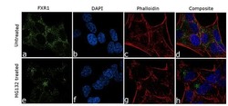
- For immunofluorescence analysis, SH-SY5Y cells were fixed and permeabilized for detection of endogenous FXR1 using Anti-FXR1 Recombinant Rabbit Monoclonal Antibody (Product # 702410, 5µg/mL) and labeled with Goat anti-Rabbit IgG (H+L) Superclonal™ Secondary Antibody, Alexa Fluor® 488 conjugate (Product # A27034, 1:2000). Nuclei (blue) is stained using SlowFade® Gold Antifade Mountant with DAPI (Product # S36938) and cytoskeletal F-actin (red) staining using Rhodamine Phalloidin (Product # R415, 1:300).Panel a-d) shows representative cells in normal growth conditions that were stained for detection and localization of FXR1 protein (green) with diffused cytoplasmic signal. Panel e-h) clearly demonstrate translocation of FXR1 to cytoplasmic stress granules in cells treated with MG132 (10 µg/mL 3h). The images were captured at 60X magnification.

Supportive validation
- Submitted by
- Invitrogen Antibodies (provider)
- Main image

- Experimental details
- RNA iImmunoprecipitation (RIP) western of FXR1 was performed on K562 cells. Antigen-antibody complexes were formed by incubating approximately 500 µg whole cell lysate with 5 µg of FXR1 monoclonal antibody (Product # 702410) rotating 60 min at RT. The immune complexes were captured on 625 µg of anti-rabbit coated Dynabeads (Product # 11204D), washed extensively, and eluted with NuPAGE™ LDS Sample Buffer (Product # NP0007). Samples were resolved onto NuPAGE™ 4-12% Bis-Tris gel (Product # NP0335BOX). Lanes 1 and 3 are input and lanes 2 and 4 are IP. Proteins were transferred to PVDF membrane (Product # IB23001). Membrane was blocked in 5% milk. Target was detected using a FXR1 monoclonal antibody (Product # 702410) at a dilution of 1:2000, followed by a 1:4000 dilution of secondary antibody. Chemiluminescent detection was performed using ECL Western Blotting Substrate (Product # 32106). Data courtesy of the Yeo lab as part of the ENCODE project (www.encodeproject.org).
