Explore
 Validate
 Learn
Learn Western blot
 Immunocytochemistry
 Immunohistochemistry
Antibody data
- Antibody Data
- Antigen structure
- References [3]
- Comments [0]
- Validations
- Western blot [1]
- Immunocytochemistry [1]
Submit
Validation data
Reference
Comment
Report error
- Product number
- HPA006964 - Provider product page

- Provider
- Atlas Antibodies
- Proper citation
- Atlas Antibodies Cat#HPA006964, RRID:AB_1856015
- Product name
- Anti-RAB7A
- Antibody type
- Polyclonal
- Description
- Polyclonal Antibody against Human RAB7A, Gene description: RAB7A, member RAS oncogene family, Alternative Gene Names: RAB7, Validated applications: WB, IHC, ICC, Uniprot ID: P51149, Storage: Store at +4°C for short term storage. Long time storage is recommended at -20°C.
- Reactivity
- Human, Mouse, Rat
- Host
- Rabbit
- Conjugate
- Unconjugated
- Isotype
- IgG
- Vial size
- 100 µl
- Concentration
- 0.2 mg/ml
- Storage
- Store at +4°C for short term storage. Long time storage is recommended at -20°C.
- Handling
- The antibody solution should be gently mixed before use.
Submitted references Differential response of placental cells to high D‐glucose and its impact on extracellular vesicle biogenesis and trafficking via small GTPase Ras‐related protein RAB‐7A
A SPLICS reporter reveals $${{{{{\boldsymbol{\alpha }}}}}}$$-synuclein regulation of lysosome-mitochondria contacts which affects TFEB nuclear translocation
Exploring the Impact of PARK2 Mutations on the Total and Mitochondrial Proteome of Human Skin Fibroblasts
Palma C, Lai A, Scholz‐Romero K, Chittoory H, Van Haeringen B, Carrion F, Handberg A, Lappas M, Lakhani S, McCart Reed A, McIntyre H, Nair S, Salomon C
Journal of Extracellular Biology 2024;3(1)
Journal of Extracellular Biology 2024;3(1)
A SPLICS reporter reveals $${{{{{\boldsymbol{\alpha }}}}}}$$-synuclein regulation of lysosome-mitochondria contacts which affects TFEB nuclear translocation
Giamogante F, Barazzuol L, Maiorca F, Poggio E, Esposito A, Masato A, Napolitano G, Vagnoni A, Calì T, Brini M
Nature Communications 2024;15(1)
Nature Communications 2024;15(1)
Exploring the Impact of PARK2 Mutations on the Total and Mitochondrial Proteome of Human Skin Fibroblasts
Zilocchi M, Colugnat I, Lualdi M, Meduri M, Marini F, Corasolla Carregari V, Moutaoufik M, Phanse S, Pieroni L, Babu M, Garavaglia B, Fasano M, Alberio T
Frontiers in Cell and Developmental Biology 2020;8
Frontiers in Cell and Developmental Biology 2020;8
No comments: Submit comment
Enhanced validation
- Submitted by
- Atlas Antibodies (provider)
- Enhanced method
- Genetic validation
- Main image
- Experimental details
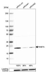
- Western blot analysis in A-431 cells transfected with control siRNA, target specific siRNA probe #1 and #2, using Anti-RAB7A antibody. Remaining relative intensity is presented. Loading control: Anti-GAPDH.
- Sample type
- Human
- Protocol
- Protocol

Supportive validation
- Submitted by
- Atlas Antibodies (provider)
- Main image
- Experimental details
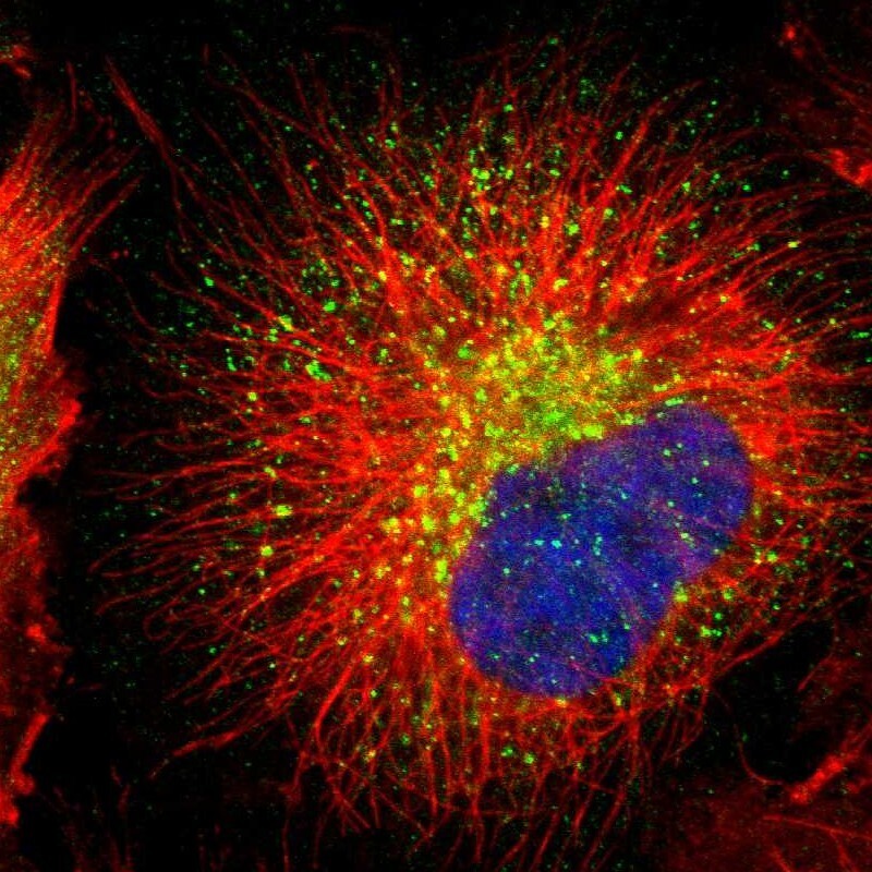
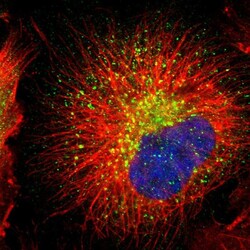
- Immunofluorescent staining of human cell line U-251 MG shows localization to lysosomes.
- Sample type
- Human