Explore
 Validate
 Learn
Learn Western blot
 Immunocytochemistry
 Immunoprecipitation
Antibody data
- Antibody Data
- Antigen structure
- References [2]
- Comments [0]
- Validations
- Immunocytochemistry [1]
- Immunohistochemistry [2]
Submit
Validation data
Reference
Comment
Report error
- Product number
- PA5-29876 - Provider product page

- Provider
- Invitrogen Antibodies
- Product name
- CHP1 Polyclonal Antibody
- Antibody type
- Polyclonal
- Antigen
- Recombinant full-length protein
- Description
- Recommended positive controls: HeLa, HepG2, mouse liver, rat liver. Predicted reactivity: Mouse (98%), Rat (98%), Zebrafish (90%), Xenopus laevis (94%), Chicken (96%), Rhesus Monkey (100%), Bovine (99%). Store product as a concentrated solution. Centrifuge briefly prior to opening the vial.
- Reactivity
- Human, Mouse, Rat
- Host
- Rabbit
- Isotype
- IgG
- Vial size
- 100 μL
- Concentration
- 1 mg/mL
- Storage
- Store at 4°C short term. For long term storage, store at -20°C, avoiding freeze/thaw cycles.
Submitted references PLS3 Overexpression Delays Ataxia in Chp1 Mutant Mice.
An orthogonal proteomic survey uncovers novel Zika virus host factors.
Janzen E, Wolff L, Mendoza-Ferreira N, Hupperich K, Delle Vedove A, Hosseinibarkooie S, Kye MJ, Wirth B
Frontiers in neuroscience 2019;13:993
Frontiers in neuroscience 2019;13:993
An orthogonal proteomic survey uncovers novel Zika virus host factors.
Scaturro P, Stukalov A, Haas DA, Cortese M, Draganova K, Płaszczyca A, Bartenschlager R, Götz M, Pichlmair A
Nature 2018 Sep;561(7722):253-257
Nature 2018 Sep;561(7722):253-257
No comments: Submit comment
Supportive validation
- Submitted by
- Invitrogen Antibodies (provider)
- Main image
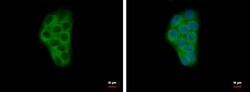
- Experimental details
- CHP1 Polyclonal Antibody detects Calcium binding protein P22 protein at cytoplasm by immunofluorescent analysis. Sample: HepG2 cells were fixed in 4% paraformaldehyde at RT for 15 min. Green: Calcium binding protein P22 protein stained by CHP1 Polyclonal Antibody (Product # PA5-29876) diluted at 1:500. Blue: Hoechst 33342 staining.

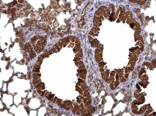
Supportive validation
- Submitted by
- Invitrogen Antibodies (provider)
- Main image

- Experimental details
- Calcium binding protein P22 antibody detects Calcium binding protein P22 protein at cytosol on mouse lung by immunohistochemical analysis. Sample: Paraffin-embedded mouse lung. Calcium binding protein P22 antibody (Product # PA5-29876) dilution: 1:500. Antigen Retrieval: EDTA based buffer, pH 8.0, 15 min.
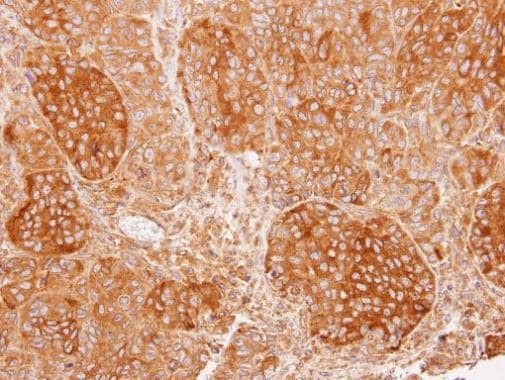
- Submitted by
- Invitrogen Antibodies (provider)
- Main image

- Experimental details
- Immunohistochemical analysis of paraffin-embedded SW480 xenograft, using Calcium binding protein P22 (Product # PA5-29876) antibody at 1:100 dilution. Antigen Retrieval: EDTA based buffer, pH 8.0, 15 min.