Explore
 Validate
 Learn
Learn Western blot
 Immunocytochemistry
Antibody data
- Antibody Data
- Antigen structure
- References [1]
- Comments [0]
- Validations
- Immunocytochemistry [2]
- Other assay [1]
Submit
Validation data
Reference
Comment
Report error
- Product number
- PA5-14363 - Provider product page

- Provider
- Invitrogen Antibodies
- Product name
- EN2 Polyclonal Antibody
- Antibody type
- Polyclonal
- Antigen
- Synthetic peptide
- Description
- This antibody is predicted to react with chicken, Xenopus and zebrafish based on sequence homology.
- Reactivity
- Human, Mouse
- Host
- Rabbit
- Isotype
- IgG
- Vial size
- 400 μL
- Concentration
- 2 mg/mL
- Storage
- Store at 4°C short term. For long term storage, store at -20°C, avoiding freeze/thaw cycles.
Submitted references Dual analysis of the murine cytomegalovirus and host cell transcriptomes reveal new aspects of the virus-host cell interface.
Juranic Lisnic V, Babic Cac M, Lisnic B, Trsan T, Mefferd A, Das Mukhopadhyay C, Cook CH, Jonjic S, Trgovcich J
PLoS pathogens 2013;9(9):e1003611
PLoS pathogens 2013;9(9):e1003611
No comments: Submit comment
Supportive validation
- Submitted by
- Invitrogen Antibodies (provider)
- Main image
- Experimental details
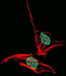
- Immunofluorescent analysis of HeLa cells stained with an EN2 polyclonal antibody (Product # PA5-14363). HeLa cells were fixed with 4% PFA (20 min), permeabilized with Triton X-100 (0.1%, 10 min), then incubated with an EN2 polyclonal antibody (Product # PA5-14363) (1:25, 1 hr at 37°C). Primary antibody was detected with fluor-conjugated donkey anti-rabbit secondary antibody (green) at 1:400 dilution for 50 min at 37°C). Actin filaments have been labeled with dye-conjugated phalloidin (red). Nuclei were counterstained with DAPI (blue) (10 µg/mL, 10 min).
- Submitted by
- Invitrogen Antibodies (provider)
- Main image

- Experimental details
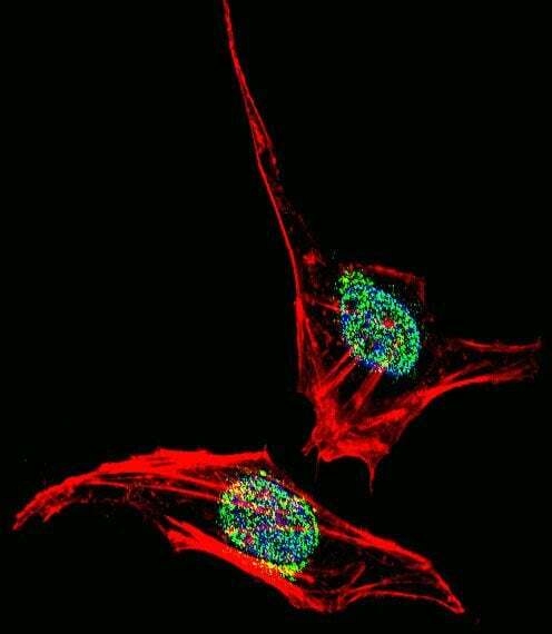
- Immunocytochemistry analysis of EN2 in HeLa cells. Samples were incubated with EN2 polyclonal antibody (Product # PA5-14363) using a dilution of 1:25 for 1 h at 37°C followed by Alexa Fluor® 488 conjugated donkey anti-rabbit at a dilution of 1:400 for 50 min at 37°C. Cells were fixed with 4% PFA (20 min) and permeabilized with Triton X-100 (0.1%, 10 min). Cytoplasmic actin was counterstained with Alexa Fluor® 555 (red) conjugated Phalloidin (7 units/mL, 1 h at 37°C). Nuclei were counterstained with DAPI (blue) (10 µg/mL, 10 min). EN2 immunoreactivity is localized to nucleus significantly and Cytoplasm weakly.

Supportive validation
- Submitted by
- Invitrogen Antibodies (provider)
- Main image

- Experimental details
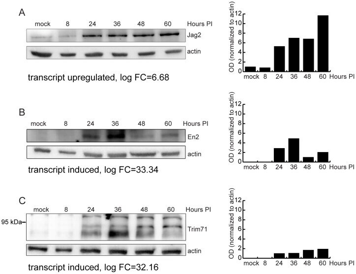
- Figure 6 Validation of RNA-Seq analysis of host genes by western blot. (A) Immunoblot analysis of MEF.K (A) or Balb/c MEF (B-C) cell lysates infected with wild-type MCMV. Cell lysates were separated by SDS-PAGE, transferred to PVDF membrane, and probed with antibody to Jag2 (A), EN2 (B) or Trim71 (C). Monoclonal antibody to actin was used as loading control. Bar charts represent relative quantification of proteins. In the case of Trim71 (C where anti-Trim71 antibody detected multiple bands, the bars show quantification of the middle band.