Explore
 Validate
 Learn
Learn Western blot
 Immunocytochemistry
Antibody data
- Antibody Data
- Antigen structure
- References [5]
- Comments [0]
- Validations
- Western blot [1]
- Immunohistochemistry [6]
Submit
Validation data
Reference
Comment
Report error
- Product number
- NB300-167 - Provider product page

- Provider
- Novus Biologicals
- Proper citation
- Novus Cat#NB300-167, RRID:AB_2187584
- Product name
- Rabbit Polyclonal SHANK1 Antibody
- Antibody type
- Polyclonal
- Description
- Immunogen affinity purified. Reacts with residues [SGPIYPGLFDIRSS] of the C-terminus of the SHANK1 protein.
- Reactivity
- Human, Mouse, Rat
- Host
- Rabbit
- Isotype
- IgG
- Vial size
- 0.1 ml
- Concentration
- 1 mg/ml
- Storage
- Store at 4C short term. Aliquot and store at -20C long term. Avoid freeze-thaw cycles.
Submitted references Transgenerational Bisphenol A Causes Deficits in Social Recognition and Alters Postsynaptic Density Genes in Mice.
USP8 Deubiquitinates SHANK3 to Control Synapse Density and SHANK3 Activity-Dependent Protein Levels.
Selective Localization of Shanks to VGLUT1-Positive Excitatory Synapses in the Mouse Hippocampus.
Effects of trace metal profiles characteristic for autism on synapses in cultured neurons.
Autistic-like behaviours and hyperactivity in mice lacking ProSAP1/Shank2.
Wolstenholme JT, Drobná Z, Henriksen AD, Goldsby JA, Stevenson R, Irvin JW, Flaws JA, Rissman EF
Endocrinology 2019 Aug 1;160(8):1854-1867
Endocrinology 2019 Aug 1;160(8):1854-1867
USP8 Deubiquitinates SHANK3 to Control Synapse Density and SHANK3 Activity-Dependent Protein Levels.
Kerrisk Campbell M, Sheng M
The Journal of neuroscience : the official journal of the Society for Neuroscience 2018 Jun 6;38(23):5289-5301
The Journal of neuroscience : the official journal of the Society for Neuroscience 2018 Jun 6;38(23):5289-5301
Selective Localization of Shanks to VGLUT1-Positive Excitatory Synapses in the Mouse Hippocampus.
Heise C, Schroeder JC, Schoen M, Halbedl S, Reim D, Woelfle S, Kreutz MR, Schmeisser MJ, Boeckers TM
Frontiers in cellular neuroscience 2016;10:106
Frontiers in cellular neuroscience 2016;10:106
Effects of trace metal profiles characteristic for autism on synapses in cultured neurons.
Hagmeyer S, Mangus K, Boeckers TM, Grabrucker AM
Neural plasticity 2015;2015:985083
Neural plasticity 2015;2015:985083
Autistic-like behaviours and hyperactivity in mice lacking ProSAP1/Shank2.
Schmeisser MJ, Ey E, Wegener S, Bockmann J, Stempel AV, Kuebler A, Janssen AL, Udvardi PT, Shiban E, Spilker C, Balschun D, Skryabin BV, Dieck St, Smalla KH, Montag D, Leblond CS, Faure P, Torquet N, Le Sourd AM, Toro R, Grabrucker AM, Shoichet SA, Schmitz D, Kreutz MR, Bourgeron T, Gundelfinger ED, Boeckers TM
Nature 2012 Apr 29;486(7402):256-60
Nature 2012 Apr 29;486(7402):256-60
No comments: Submit comment
Supportive validation
- Submitted by
- Novus Biologicals (provider)
- Main image
- Experimental details
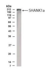
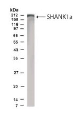
- Western Blot: SHANK1 Antibody [NB300-167] - Shank1a in rat brain lysate using NB300-167 at 1:500 dilution.
Supportive validation
- Submitted by
- Novus Biologicals (provider)
- Main image
- Experimental details
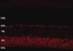
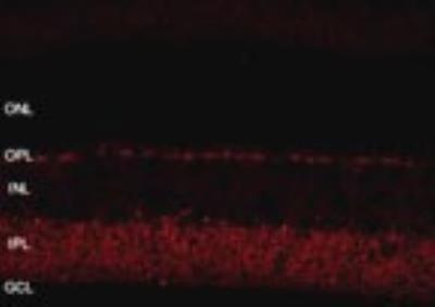
- Immunohistochemistry: SHANK1 Antibody [NB300-167] - Retinal tissue using NB300-167 at 1:800 dilution. Images are 2048 X 2048, 12 bit, collected with a 40X water objective, 10 stacks 0.55 um per stack.
- Submitted by
- Novus Biologicals (provider)
- Main image
- Experimental details
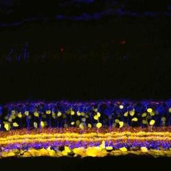
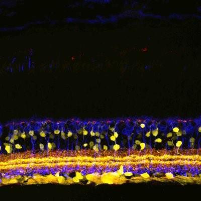
- Immunohistochemistry: SHANK1 Antibody [NB300-167] - Mouse retina using NB300-167.
- Submitted by
- Novus Biologicals (provider)
- Main image
- Experimental details
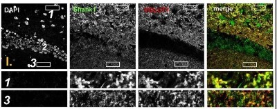
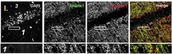
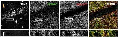
- Immunohistochemistry: SHANK1 Antibody [NB300-167] - Colocalization of Shank puncta with VGLUT1 puncta in the stratum radiatum of the CA1. confocal immunofluorescence stainings of coronal sections from wild-type mice probed with the Shank1 (white; green in merge) and VGLUT1 (white; red in merge) antibodies. The upper row (large squares) shows the enlarged region (3 = SO; 2 = SP; 1 = SR; scale bar = 100 um), the bottom row (small rectangles) shows a further enlargement (indicated in upper row by white rectangle) in the SR (scale bar = 5 um). Image collected and cropped by CiteAb from the following publication (http://journal.frontiersin.org/Article/10.3389/fncel.2016.00106/abstract) licensed under a CC-BY licence.
- Submitted by
- Novus Biologicals (provider)
- Main image

- Experimental details
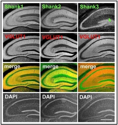
- Immunohistochemistry: SHANK1 Antibody [NB300-167] - Synaptic distribution of Shank1/2 and somato-synaptic distribution of Shank3 in the mouse hippocampus, codistribution with VGLUT1, no codistribution with VGLUT2. 5x magnification of hippocampus. Immunofluorescence stainings of coronal sections from wild-type mice probed with Shank1-3 (white; green in merge) and VGLUT1 (white; red in merge) antibodies; green arrow points toward the intragranular mossy fibers where there is a prominent synaptic stain of Shank3; scale bar =300 um. Image collected and cropped by CiteAb from the following publication (http://journal.frontiersin.org/Article/10.3389/fncel.2016.00106/abstract) licensed under a CC-BY licence.
- Submitted by
- Novus Biologicals (provider)
- Main image
- Experimental details
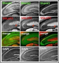
- Immunohistochemistry: SHANK1 Antibody [NB300-167] - Synaptic distribution of Shank1/2 and somato-synaptic distribution of Shank3 in the mouse hippocampus, codistribution with VGLUT1, no codistribution with VGLUT2. Immunofluorescence stainings of coronal sections from wild-type mice probed with Shank1-3 (white; green in merge) and VGLUT2 (white; red in merge) antibodies; green arrow points toward the intragranular mossy fibers where there is a prominent synaptic stain of Shank3. Red arrow points toward the VGLUT2-band of the DG; scale bar = 300 um. Image collected and cropped by CiteAb from the following publication (http://journal.frontiersin.org/Article/10.3389/fncel.2016.00106/abstract) licensed under a CC-BY licence.

- Submitted by
- Novus Biologicals (provider)
- Main image
- Experimental details
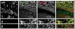
- Immunohistochemistry: SHANK1 Antibody [NB300-167] - Colocalization of Shank puncta with VGLUT1 puncta in the intragranular mossy fibers and the stratum moleculare of the dentate gyrus. Confocal immunofluorescence stainings of coronal sections from wild-type mice probed with the Shank1 (white; green in merge) and VGLUT1 (white; red in merge) antibodies. The upper rows (large squares) show the enlarged region (3 = SM; 2 = SG; 1 = PL; scale bar = 100 um), the bottom rows (small rectangles) show further enlargements (indicated in upper row by white rectangles) in the PL (1, top row) and SM (3, bottom row) (scale bar = 5 um). Image collected and cropped by CiteAb from the following publication (http://journal.frontiersin.org/Article/10.3389/fncel.2016.00106/abstract) licensed under a CC-BY licence.