 Explore
 Validate
 Learn
LearnPA5-51933
antibody from Invitrogen Antibodies
Targeting: SYNE2
DKFZP434H2235, KIAA1011, Nesp2, Nesprin-2, NUA, NUANCE, SYNE-2
 Immunocytochemistry
Antibody data
- Antibody Data
- Antigen structure
- References [1]
- Comments [0]
- Validations
- Immunocytochemistry [1]
- Immunohistochemistry [5]
- Other assay [1]
Submit
Validation data
Reference
Comment
Report error
- Product number
- PA5-51933 - Provider product page

- Provider
- Invitrogen Antibodies
- Product name
- Nesprin 2 Polyclonal Antibody
- Antibody type
- Polyclonal
- Antigen
- Recombinant protein fragment
- Description
- Immunogen sequence: NVLNDAYENL TRYKEAVTRA VESITSLEAI IIPYRVDVGN PEESLEMPLR KQEELESTVA RIQDLTEKLG MISSPEAKLQ LQYTLQELVS KNSAMKEAFK AQETEAE Highest antigen sequence identity to the following orthologs: Mouse - 56%, Rat - 29%.
- Reactivity
- Human
- Host
- Rabbit
- Isotype
- IgG
- Vial size
- 100 μL
- Concentration
- 0.3 mg/mL
- Storage
- Store at 4°C short term. For long term storage, store at -20°C, avoiding freeze/thaw cycles.
Submitted references Lamin A-mediated nuclear lamina integrity is required for proper ciliogenesis.
Fan JR, You LR, Wang WJ, Huang WS, Chu CT, Chi YH, Chen HC
EMBO reports 2020 Oct 5;21(10):e49680
EMBO reports 2020 Oct 5;21(10):e49680
No comments: Submit comment
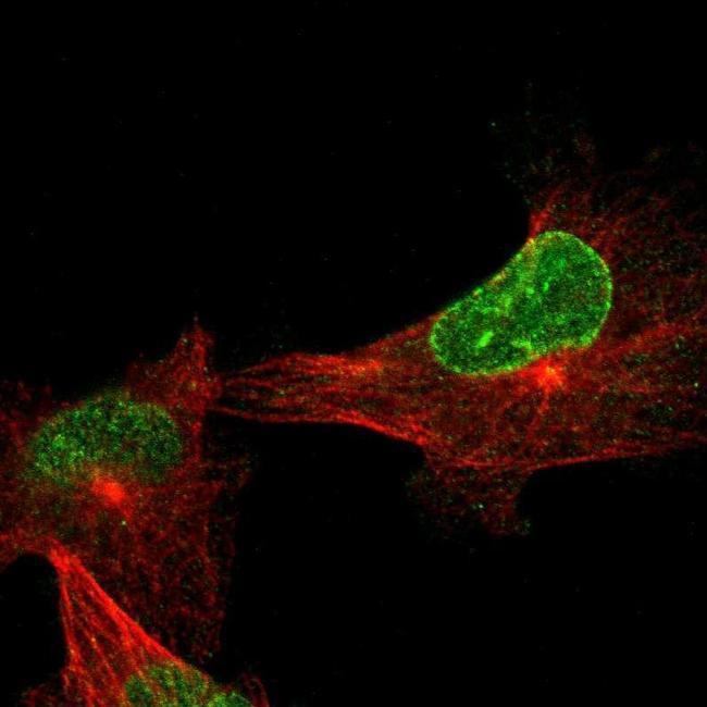
Supportive validation
- Submitted by
- Invitrogen Antibodies (provider)
- Main image

- Experimental details
- Immunofluorescent staining of Nesprin 2 in human cell line U-251 MG shows positivity in nucleus & nuclear membrane. Samples were probed using a Nesprin 2 Polyclonal Antibody (Product # PA5-51933).
Supportive validation
- Submitted by
- Invitrogen Antibodies (provider)
- Main image
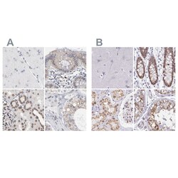
- Experimental details
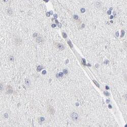
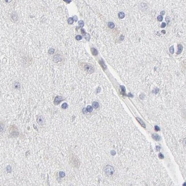
- Immunohistochemical staining of Nesprin 2 in human cerebral cortex, colon, kidney and testis using Nesprin 2 Polyclonal Antibody (Product # PA5-51933) (A) shows similar protein distribution across tissues to an independent Nesprin 2 Polyclonal Antibody (B).

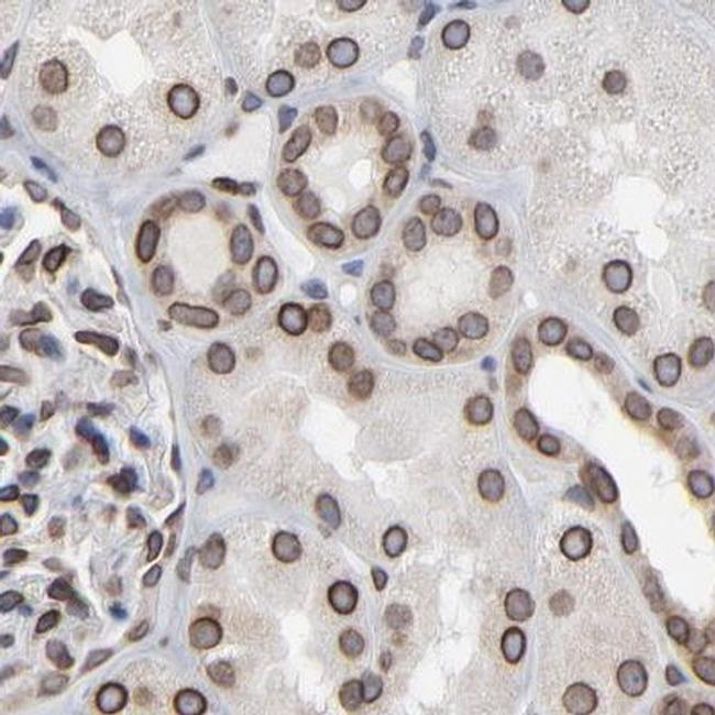
- Submitted by
- Invitrogen Antibodies (provider)
- Main image
- Experimental details
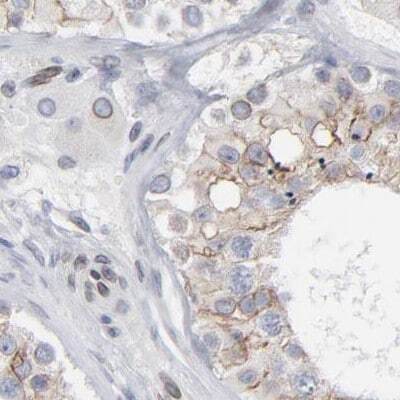
- Immunohistochemical staining of Nesprin 2 in human kidney using Nesprin 2 Polyclonal Antibody (Product # PA5-51933).
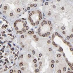
- Submitted by
- Invitrogen Antibodies (provider)
- Main image
- Experimental details
- Immunohistochemical staining of Nesprin 2 in human cerebral cortex using Nesprin 2 Polyclonal Antibody (Product # PA5-51933).
- Submitted by
- Invitrogen Antibodies (provider)
- Main image

- Experimental details
- Immunohistochemical staining of Nesprin 2 in human colon using Nesprin 2 Polyclonal Antibody (Product # PA5-51933).

- Submitted by
- Invitrogen Antibodies (provider)
- Main image

- Experimental details
- Immunohistochemical staining of Nesprin 2 in human testis using Nesprin 2 Polyclonal Antibody (Product # PA5-51933).
Supportive validation
- Submitted by
- Invitrogen Antibodies (provider)
- Main image
- Experimental details
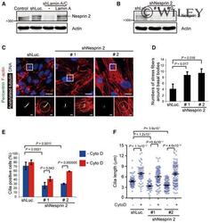
- Impaired ciliogenesis by depletion of lamin A/C may partially result from nesprin 2 suppression A An equal amount of whole-cell lysates from RPE cells or those expressing shLamin A/C or luciferase (shLuc) was analyzed by the immunoblotting with antibodies as indicated. B-D RPE cells were infected with lentiviruses expressing shRNAs to nesprin 2 (shNesprine 2, clone #1, and clone #2) or luciferase (shLuc) as a control. An equal amount of whole-cell lysates was analyzed by immunoblotting with antibodies as indicated (B). The cells were serum-starved for 48 h and then stained for acetylated tubulin (white), pericentrin (green), F-actin (red), and DNA (blue). The representative images are shown (C) The left insets show the merged signals of acetylated tubulin and pericentrin. Scale bars, 20 or 2 mum (magnified images). The numbers of F-actin stress fibers within 60 mum 2 around the basal bodies (as illustrated by the circle) were measured (D, n >= 53). E, F RPE cells expressing shNesprine 2 or shLuc were serum-starved for 32 h and then treated with (+) or without (-) Cyto D for another 16 h. The percentage of the cells with cilia in the total counted cells (E, n >= 197) and the length of cilia (F, n >= 52) were measured. Data information: In D and E, values (means +- SD) are from three independent experiments. In F, values (means +- SEM) are from three independent experiments. Statistical significance of differences is assessed with Student's t -test.
