Explore
 Validate
 Learn
Learn Western blot
 Immunohistochemistry
Antibody data
- Antibody Data
- Antigen structure
- References [4]
- Comments [0]
- Validations
- Immunohistochemistry [1]
Submit
Validation data
Reference
Comment
Report error
- Product number
- PA3-021 - Provider product page

- Provider
- Invitrogen Antibodies
- Product name
- GPR32 Polyclonal Antibody
- Antibody type
- Polyclonal
- Antigen
- Other
- Description
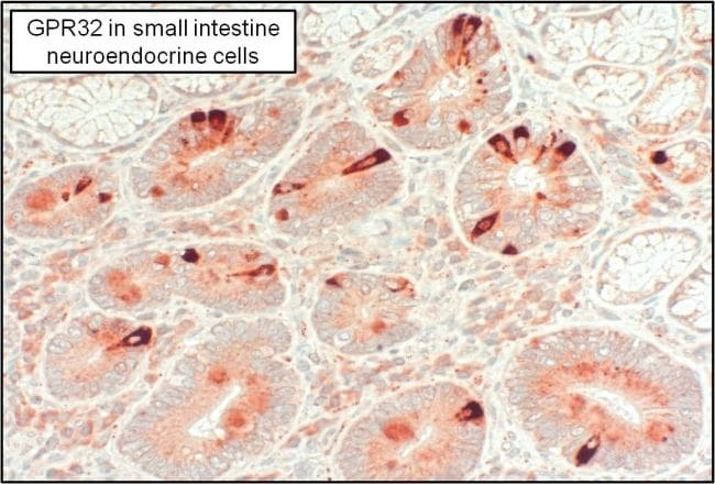
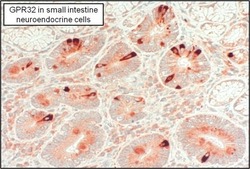
- IHC (P) analysis shows positive staining of GPR32 in human small intestine tissue neuroendocrine cells. WB analysis shows GPR32 in glycoprotein-enriched fractions from GPR32 overexpressing 293 cells. Additional unknown bands at ~30kD and ~130kD were also detected. While GPR32 has a theoretical MW of ~ 40kD, the observed band in a WB runs at ~60kD due to enrichment of the glycosylated receptor.
- Reactivity
- Human
- Host
- Rabbit
- Isotype
- IgG
- Vial size
- 100 μL
- Concentration
- Conc. Not Determined
- Storage
- -20°C
Submitted references Passive and active inclusion of host proteins in human immunodeficiency virus type 1 gag particles during budding at the plasma membrane.
Differential display implicates cyclophilin A in adult cortical plasticity.
Interaction of the retinoblastoma gene product, RB, with cyclophilin A negatively affects cyclosporin-inhibited NFAT signaling.
The hydrophobic pocket of cyclophilin is the binding site for the human immunodeficiency virus type 1 Gag polyprotein.
Hammarstedt M, Garoff H
Journal of virology 2004 Jun;78(11):5686-97
Journal of virology 2004 Jun;78(11):5686-97
Differential display implicates cyclophilin A in adult cortical plasticity.
Arckens L, Van der Gucht E, Van den Bergh G, Massie A, Leysen I, Vandenbussche E, Eysel UT, Huybrechts R, Vandesande F
The European journal of neuroscience 2003 Jul;18(1):61-75
The European journal of neuroscience 2003 Jul;18(1):61-75
Interaction of the retinoblastoma gene product, RB, with cyclophilin A negatively affects cyclosporin-inhibited NFAT signaling.
Cui Y, Mirkia K, Florence Fu YH, Zhu L, Yokoyama KK, Chiu R
Journal of cellular biochemistry 2002;86(4):630-41
Journal of cellular biochemistry 2002;86(4):630-41
The hydrophobic pocket of cyclophilin is the binding site for the human immunodeficiency virus type 1 Gag polyprotein.
Braaten D, Ansari H, Luban J
Journal of virology 1997 Mar;71(3):2107-13
Journal of virology 1997 Mar;71(3):2107-13
No comments: Submit comment
Supportive validation
- Submitted by
- Invitrogen Antibodies (provider)
- Main image
- Experimental details
- Immunohistochemistry analysis of GPR32 was performed on neuroendocrine cells in human small intestine tissue. To expose target proteins, antigen retrieval was performed by microwaving tissues for 20 minutes in 10mM sodium citrate buffer (pH 6.0). Tissue slides were probed with a GPR32 polyclonal antibody (Product # PA3-021) at a dilution of 1:3000, overnight at 4C in a humidified chamber. Tissues were washed, and detection was performed using an ABC kit composed of biotinylated goat anti-rabbit IgG, peroxidase-conjugated avidin, and 3-amino-9-ethylcarbazole (AEC) substrate in acetate buffer. Tissues were counterstained with hematoxylin and dehydrated to prep for mounting.