Explore
 Validate
 Learn
Learn Immunocytochemistry
 Immunohistochemistry
Antibody data
- Antibody Data
- Antigen structure
- References [29]
- Comments [0]
- Validations
- Immunocytochemistry [2]
- Immunohistochemistry [1]
Submit
Validation data
Reference
Comment
Report error
- Product number
- HPA008346 - Provider product page

- Provider
- Atlas Antibodies
- Proper citation
- Atlas Antibodies Cat#HPA008346, RRID:AB_1080462
- Product name
- Anti-SUN1
- Antibody type
- Polyclonal
- Description
- Polyclonal Antibody against Human SUN1, Gene description: Sad1 and UNC84 domain containing 1, Alternative Gene Names: FLJ12407, KIAA0810, UNC84A, Validated applications: IHC, ICC, Uniprot ID: O94901, Storage: Store at +4°C for short term storage. Long time storage is recommended at -20°C.
- Reactivity
- Human
- Host
- Rabbit
- Conjugate
- Unconjugated
- Isotype
- IgG
- Vial size
- 100 µl
- Concentration
- 0.2 mg/ml
- Storage
- Store at +4°C for short term storage. Long time storage is recommended at -20°C.
- Handling
- The antibody solution should be gently mixed before use.
Submitted references Non-random spatial organization of telomeres varies during the cell cycle and requires LAP2 and BAF
SUN1 inhibits osteogenesis and promotes adipogenesis of human adipose-derived stem cells by regulating α-tubulin and CD36 expression.
Nucleocytoplasmic transport rates are regulated by cellular processes that modulate GTP availability.
The LINC complex ensures accurate centrosome positioning during prophase.
Activation of endoplasmic reticulum stress in premature aging via the inner nuclear membrane protein SUN2
Histone methyltransferase SUV39H1 regulates the Golgi complex via the nuclear envelope-spanning LINC complex
A membrane-sensing mechanism links lipid metabolism to protein degradation at the nuclear envelope.
SUN2 Modulates the Propagation of HSV-1
Emerin self-assembly and nucleoskeletal coupling regulate nuclear envelope mechanics against stress
Lamin A and the LINC complex act as potential tumor suppressors in Ewing Sarcoma
Inner Nuclear Membrane Protein, SUN1, is Required for Cytoskeletal Force Generation and Focal Adhesion Maturation
Baricitinib, a JAK-STAT Inhibitor, Reduces the Cellular Toxicity of the Farnesyltransferase Inhibitor Lonafarnib in Progeria Cells
Parental genome unification is highly error-prone in mammalian embryos
The SUN2-nesprin-2 LINC complex and KIF20A function in the Golgi dispersal
Progerin impairs 3D genome organization and induces fragile telomeres by limiting the dNTP pools
Nuclear Pore Complexes Cluster in Dysmorphic Nuclei of Normal and Progeria Cells during Replicative Senescence
Nesprin-1-alpha2 associates with kinesin at myotube outer nuclear membranes, but is restricted to neuromuscular junction nuclei in adult muscle
Nuclear Envelope Transmembrane Proteins in Myotonic Dystrophy Type 1
Autophagic Removal of Farnesylated Carboxy-Terminal Lamin Peptides
SUN1 splice variants, SUN1_888, SUN1_785, and predominant SUN1_916, variably function in directional cell migration
Progerin impairs chromosome maintenance by depleting CENP-F from metaphase kinetochores in Hutchinson-Gilford progeria fibroblasts
Muscular Dystrophy-Associated SUN1 and SUN2 Variants Disrupt Nuclear-Cytoskeletal Connections and Myonuclear Organization
A mammalian KASH domain protein coupling meiotic chromosomes to the cytoskeleton
Systematic validation of antibody binding and protein subcellular localization using siRNA and confocal microscopy
Human Telomeres Are Tethered to the Nuclear Envelope during Postmitotic Nuclear Assembly
Samp1 is functionally associated with the LINC complex and A-type lamina networks
Induction of a massive endoplasmic reticulum and perinuclear space expansion by expression of lamin B receptor mutants and the related sterol reductases TM7SF2 and DHCR7.
Membrane Insertion of the Pleckstrin Homology Domain Variable Loop 1 Is Critical for Dynamin-catalyzed Vesicle Scission
Keller D, Stinus S, Umlauf D, Gourbeyre E, Biot E, Olivier N, Mahou P, Beaurepaire E, Andrey P, Crabbe L
iScience 2024;27(4):109343
iScience 2024;27(4):109343
SUN1 inhibits osteogenesis and promotes adipogenesis of human adipose-derived stem cells by regulating α-tubulin and CD36 expression.
Fan T, Zhu J, Liu W, Qu R, Khan AU, Shi Y, Liu J, Zhou Z, Xu C, Dai J, Ouyang J
Journal of cellular and molecular medicine 2024 Oct;28(19):e70143
Journal of cellular and molecular medicine 2024 Oct;28(19):e70143
Nucleocytoplasmic transport rates are regulated by cellular processes that modulate GTP availability.
Scott KL, Halfmann CT, Hoefakker AD, Purkayastha P, Wang TC, Lele TP, Roux KJ
The Journal of cell biology 2024 Jul 1;223(7)
The Journal of cell biology 2024 Jul 1;223(7)
The LINC complex ensures accurate centrosome positioning during prophase.
Lima JT, Pereira AJ, Ferreira JG
Life science alliance 2024 Apr;7(4)
Life science alliance 2024 Apr;7(4)
Birks S, Howard S, Wright C, O’Rourke C, Day E, Lamb A, Walsdorf J, Lau A, Thompson W, Uzer G
2024
2024
Activation of endoplasmic reticulum stress in premature aging via the inner nuclear membrane protein SUN2
Vidak S, Serebryannyy L, Pegoraro G, Misteli T
Cell Reports 2023;42(5):112534
Cell Reports 2023;42(5):112534
Histone methyltransferase SUV39H1 regulates the Golgi complex via the nuclear envelope-spanning LINC complex
Roy A, Nishino M, Imaizumi H, Yokoyama Y, Katahira J, Kimura H, Matsuura N, Matsumura M
PLOS ONE 2023;18(7):e0283490
PLOS ONE 2023;18(7):e0283490
A membrane-sensing mechanism links lipid metabolism to protein degradation at the nuclear envelope.
Lee S, Carrasquillo Rodrı Guez JW, Merta H, Bahmanyar S
The Journal of cell biology 2023 Sep 4;222(9)
The Journal of cell biology 2023 Sep 4;222(9)
SUN2 Modulates the Propagation of HSV-1
Cruz-Palomar K, Hawkins J, Vandal C, Quenneville J, Gagnon É, Lippé R, Sandri-Goldin R
Journal of Virology 2022;96(9)
Journal of Virology 2022;96(9)
Emerin self-assembly and nucleoskeletal coupling regulate nuclear envelope mechanics against stress
Fernandez A, Bautista M, Wu L, Pinaud F
Journal of Cell Science 2022;135(6)
Journal of Cell Science 2022;135(6)
Lamin A and the LINC complex act as potential tumor suppressors in Ewing Sarcoma
Chiarini F, Paganelli F, Balestra T, Capanni C, Fazio A, Manara M, Landuzzi L, Petrini S, Evangelisti C, Lollini P, Martelli A, Lattanzi G, Scotlandi K
Cell Death & Disease 2022;13(4)
Cell Death & Disease 2022;13(4)
Inner Nuclear Membrane Protein, SUN1, is Required for Cytoskeletal Force Generation and Focal Adhesion Maturation
Ueda N, Maekawa M, Matsui T, Deguchi S, Takata T, Katahira J, Higashiyama S, Hieda M
Frontiers in Cell and Developmental Biology 2022;10
Frontiers in Cell and Developmental Biology 2022;10
Baricitinib, a JAK-STAT Inhibitor, Reduces the Cellular Toxicity of the Farnesyltransferase Inhibitor Lonafarnib in Progeria Cells
Arnold R, Vehns E, Randl H, Djabali K
International Journal of Molecular Sciences 2021;22(14):7474
International Journal of Molecular Sciences 2021;22(14):7474
Parental genome unification is highly error-prone in mammalian embryos
Cavazza T, Takeda Y, Politi A, Aushev M, Aldag P, Baker C, Choudhary M, Bucevičius J, Lukinavičius G, Elder K, Blayney M, Lucas-Hahn A, Niemann H, Herbert M, Schuh M
Cell 2021;184(11):2860-2877.e22
Cell 2021;184(11):2860-2877.e22
The SUN2-nesprin-2 LINC complex and KIF20A function in the Golgi dispersal
Hieda M, Matsumoto T, Isobe M, Kurono S, Yuka K, Kametaka S, Wang J, Chi Y, Kameda K, Kimura H, Matsuura N, Matsuura S
Scientific Reports 2021;11(1)
Scientific Reports 2021;11(1)
Progerin impairs 3D genome organization and induces fragile telomeres by limiting the dNTP pools
Kychygina A, Dall’Osto M, Allen J, Cadoret J, Piras V, Pickett H, Crabbe L
Scientific Reports 2021;11(1)
Scientific Reports 2021;11(1)
Nuclear Pore Complexes Cluster in Dysmorphic Nuclei of Normal and Progeria Cells during Replicative Senescence
Röhrl J, Arnold R, Djabali K
Cells 2021;10(1):153
Cells 2021;10(1):153
Nesprin-1-alpha2 associates with kinesin at myotube outer nuclear membranes, but is restricted to neuromuscular junction nuclei in adult muscle
Holt I, Fuller H, Lam L, Sewry C, Shirran S, Zhang Q, Shanahan C, Morris G
Scientific Reports 2019;9(1)
Scientific Reports 2019;9(1)
Nuclear Envelope Transmembrane Proteins in Myotonic Dystrophy Type 1
Hintze S, Knaier L, Limmer S, Schoser B, Meinke P
Frontiers in Physiology 2018;9
Frontiers in Physiology 2018;9
Autophagic Removal of Farnesylated Carboxy-Terminal Lamin Peptides
Lu X, Djabali K
Cells 2018;7(4):33
Cells 2018;7(4):33
SUN1 splice variants, SUN1_888, SUN1_785, and predominant SUN1_916, variably function in directional cell migration
Nishioka Y, Imaizumi H, Imada J, Katahira J, Matsuura N, Hieda M
Nucleus 2016;7(6):572-584
Nucleus 2016;7(6):572-584
Progerin impairs chromosome maintenance by depleting CENP-F from metaphase kinetochores in Hutchinson-Gilford progeria fibroblasts
Eisch V, Lu X, Gabriel D, Djabali K
Oncotarget 2016;7(17):24700-24718
Oncotarget 2016;7(17):24700-24718
Muscular Dystrophy-Associated SUN1 and SUN2 Variants Disrupt Nuclear-Cytoskeletal Connections and Myonuclear Organization
Cox G, Meinke P, Mattioli E, Haque F, Antoku S, Columbaro M, Straatman K, Worman H, Gundersen G, Lattanzi G, Wehnert M, Shackleton S
PLoS Genetics 2014;10(9):e1004605
PLoS Genetics 2014;10(9):e1004605
A mammalian KASH domain protein coupling meiotic chromosomes to the cytoskeleton
Horn H, Kim D, Wright G, Wong E, Stewart C, Burke B, Roux K
Journal of Cell Biology 2013;202(7):1023-1039
Journal of Cell Biology 2013;202(7):1023-1039
Systematic validation of antibody binding and protein subcellular localization using siRNA and confocal microscopy
Stadler C, Hjelmare M, Neumann B, Jonasson K, Pepperkok R, Uhlén M, Lundberg E
Journal of Proteomics 2012;75(7):2236-2251
Journal of Proteomics 2012;75(7):2236-2251
Human Telomeres Are Tethered to the Nuclear Envelope during Postmitotic Nuclear Assembly
Crabbe L, Cesare A, Kasuboski J, Fitzpatrick J, Karlseder J
Cell Reports 2012;2(6):1521-1529
Cell Reports 2012;2(6):1521-1529
Samp1 is functionally associated with the LINC complex and A-type lamina networks
Gudise S, Figueroa R, Lindberg R, Larsson V, Hallberg E
Journal of Cell Science 2011;124(12):2077-2085
Journal of Cell Science 2011;124(12):2077-2085
Induction of a massive endoplasmic reticulum and perinuclear space expansion by expression of lamin B receptor mutants and the related sterol reductases TM7SF2 and DHCR7.
Zwerger M, Kolb T, Richter K, Karakesisoglou I, Herrmann H
Molecular biology of the cell 2010 Jan 15;21(2):354-68
Molecular biology of the cell 2010 Jan 15;21(2):354-68
Membrane Insertion of the Pleckstrin Homology Domain Variable Loop 1 Is Critical for Dynamin-catalyzed Vesicle Scission
Ramachandran R, Pucadyil T, Liu Y, Acharya S, Leonard M, Lukiyanchuk V, Schmid S
Molecular Biology of the Cell 2009 November;20(22):4630-4639
Molecular Biology of the Cell 2009 November;20(22):4630-4639
No comments: Submit comment
Enhanced validation
Supportive validation
- Submitted by
- 55af80e3e0991
- Enhanced method
- Genetic validation
- Main image
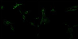
- Experimental details
- Confocal images of immunofluorescently stained human U-2 OS cells.The protein SUN1 is shown in green. The image to the left show cells transfected with control siRNA and the image to the right show cells where SUN1 has been downregulated with specific siRNA.
- Sample type
- U-2 OS cells
- Primary Ab dilution
- 1:59
- Secondary Ab
- Secondary Ab
- Secondary Ab dilution
- 1:800
- Knockdown/Genetic Approaches Application
- Immunocytochemistry
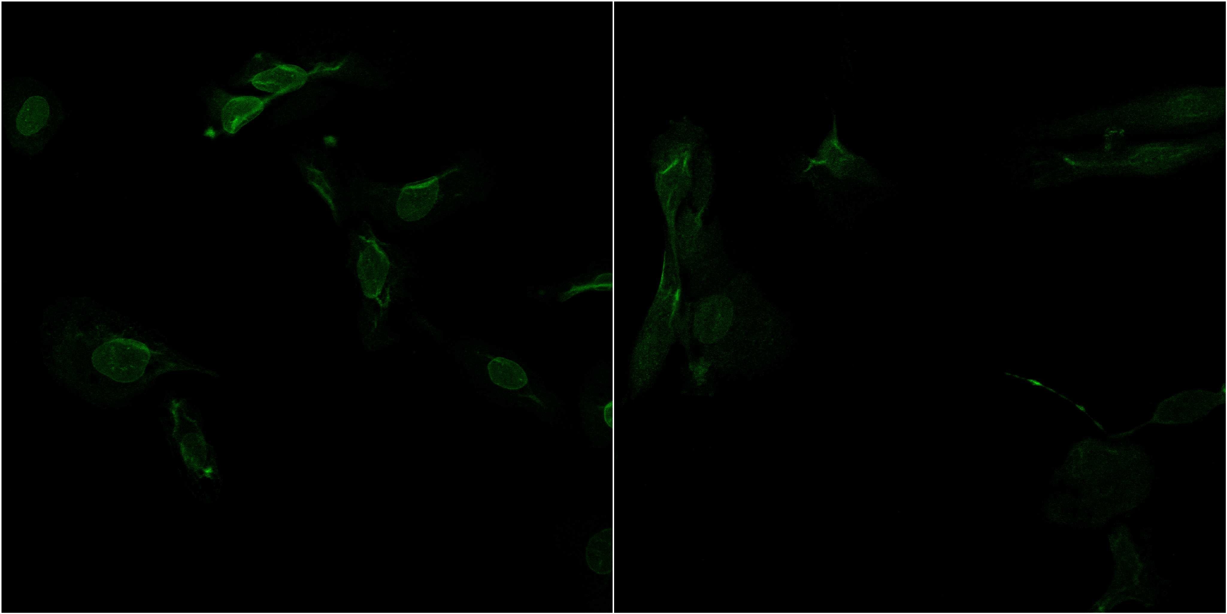
Supportive validation
- Submitted by
- Atlas Antibodies (provider)
- Main image
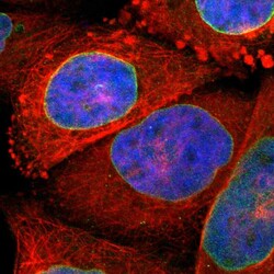
- Experimental details
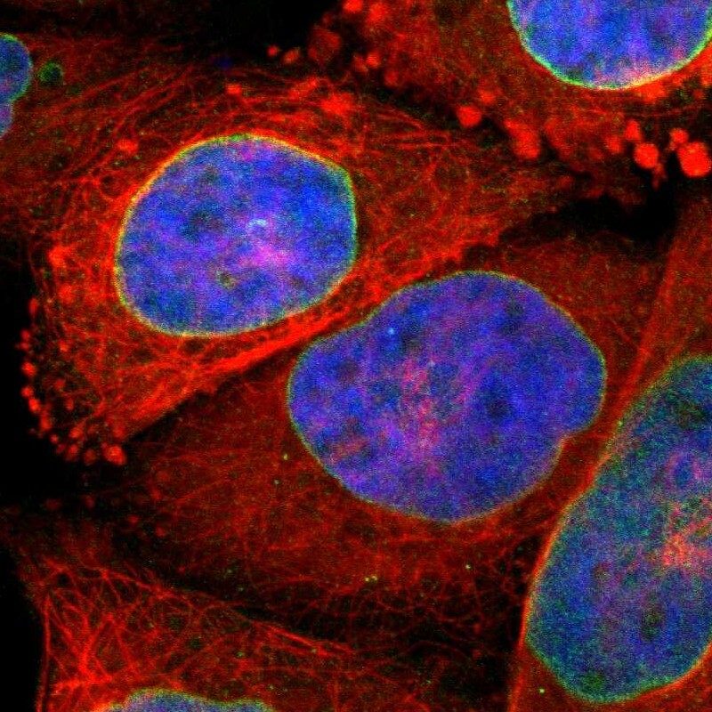
- Immunofluorescent staining of human cell line A-431 shows positivity in nuclear membrane.
- Sample type
- Human
Supportive validation
- Submitted by
- Atlas Antibodies (provider)
- Enhanced method
- Orthogonal validation
- Main image
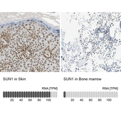
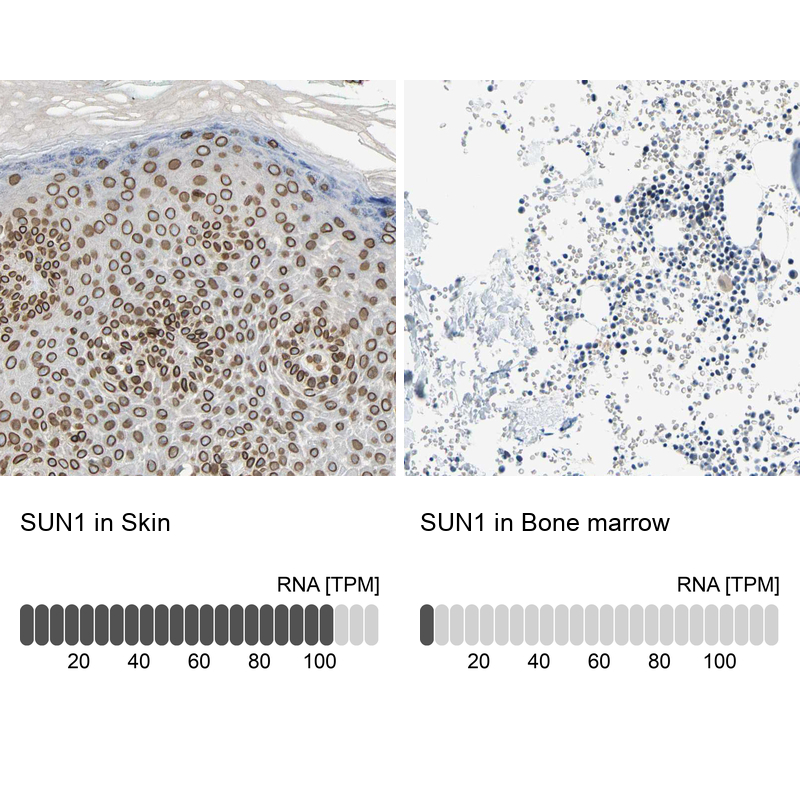
- Experimental details
- Immunohistochemistry analysis in human skin and bone marrow tissues using HPA008346 antibody. Corresponding SUN1 RNA-seq data are presented for the same tissues.
- Sample type
- Human
- Protocol
- Protocol