Explore
 Validate
 Learn
Learn Western blot
 Immunoprecipitation
Antibody data
- Antibody Data
- Antigen structure
- References [2]
- Comments [0]
- Validations
- Western blot [3]
- Immunocytochemistry [2]
- Immunohistochemistry [2]
Submit
Validation data
Reference
Comment
Report error
- Product number
- GTX101540 - Provider product page

- Provider
- GeneTex
- Proper citation
- GeneTex Cat#GTX101540, RRID:AB_1950950
- Product name
- Musashi 1 antibody [N3C3]
- Antibody type
- Polyclonal
- Reactivity
- Human, Mouse
- Host
- Rabbit
Submitted references Resources for the Comprehensive Discovery of Functional RNA Elements.
Lin28a regulates neuronal differentiation and controls miR-9 production.
Sundararaman B, Zhan L, Blue SM, Stanton R, Elkins K, Olson S, Wei X, Van Nostrand EL, Pratt GA, Huelga SC, Smalec BM, Wang X, Hong EL, Davidson JM, Lécuyer E, Graveley BR, Yeo GW
Molecular cell 2016 Mar 17;61(6):903-13
Molecular cell 2016 Mar 17;61(6):903-13
Lin28a regulates neuronal differentiation and controls miR-9 production.
Nowak JS, Choudhury NR, de Lima Alves F, Rappsilber J, Michlewski G
Nature communications 2014 Apr 11;5:3687
Nature communications 2014 Apr 11;5:3687
No comments: Submit comment
Supportive validation
- Submitted by
- GeneTex (provider)
- Main image
- Experimental details
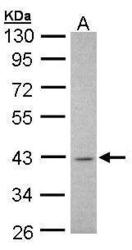
- Sample (30 ?g of whole cell lysate)A: H129910% SDS PAGEGTX101540 diluted at 1:1000The HRP-conjugated anti-rabbit IgG antibody (GTX213110-01) was used to detect the primary antibody.

- Submitted by
- GeneTex (provider)
- Main image
- Experimental details
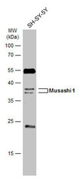
- Whole cell extract (30 ?g) was separated by 12% SDS-PAGE, and the membrane was blotted with Musashi 1 antibody [N3C3] (GTX101540) diluted at 1:1000. The HRP-conjugated anti-rabbit IgG antibody (GTX213110-01) was used to detect the primary antibody.
- Submitted by
- GeneTex (provider)
- Main image
- Experimental details
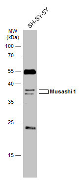
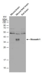
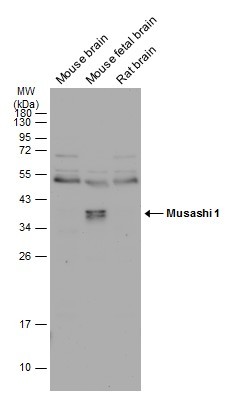
- Various tissue extracts (50 ?g) were separated by 12% SDS-PAGE, and the membrane was blotted with Musashi 1 antibody [N3C3] (GTX101540) diluted at 1:1000. The HRP-conjugated anti-rabbit IgG antibody (GTX213110-01) was used to detect the primary antibody.
Supportive validation
- Submitted by
- GeneTex (provider)
- Main image

- Experimental details
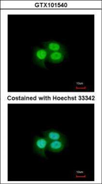
- Immunofluorescence analysis of paraformaldehyde-fixed A549, using MSI1(GTX101540) antibody at 1:200 dilution.
- Submitted by
- GeneTex (provider)
- Main image
- Experimental details
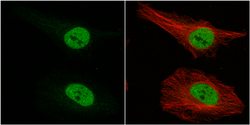
- MSI1 antibody [N3C3] detects MSI1 protein at nucleus by immunofluorescent analysis.Sample: HeLa cells were fixed in 4% paraformaldehyde at RT for 15 min.Green: MSI1 protein stained by MSI1 antibody [N3C3] (GTX101540) diluted at 1:500.Red: alpha Tubulin, a cytoskeleton marker, stained by alpha Tubulin antibody [GT114] (GTX628802) diluted at 1:1000.

Supportive validation
- Submitted by
- GeneTex (provider)
- Main image
- Experimental details
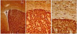
- MSI1 antibody [N3C3] detects MSI1 protein on adult mouse brain by immunohistochemical analysis. Sample: Frozen section of adult mouse brain. MSI1 antibody [N3C3] (GTX101540) diluted at 1:500.
- Submitted by
- GeneTex (provider)
- Main image

- Experimental details
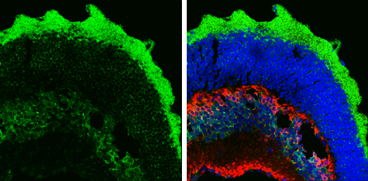
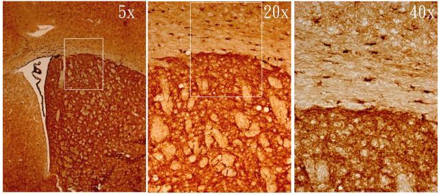
- Musashi 1 antibody [N3C3] detects Musashi 1 protein by immunohistochemical analysis.Sample: Frozen sectioned adult mouse retina. Green: Musashi 1 protein stained by Musashi 1 antibody [N3C3] (GTX101540) diluted at 1:250.Red: Protein kinase C alpha staining.Blue: Fluoroshield with DAPI (GTX30920).