Explore
 Validate
 Learn
Learn Western blot
 Immunoprecipitation
Antibody data
- Antibody Data
- Antigen structure
- References [0]
- Comments [0]
- Validations
- Western blot [6]
- Immunocytochemistry [3]
- Immunohistochemistry [2]
Submit
Validation data
Reference
Comment
Report error
- Product number
- PA5-27449 - Provider product page

- Provider
- Invitrogen Antibodies
- Product name
- MSI1 Polyclonal Antibody
- Antibody type
- Polyclonal
- Antigen
- Recombinant protein fragment
- Description
- Recommended positive controls: SH-SY-5Y, mouse fetal brain. Predicted reactivity: Mouse (99%), Rat (100%), Xenopus laevis (81%), Pig (98%), Bovine (100%). Store product as a concentrated solution. Centrifuge briefly prior to opening the vial.
- Reactivity
- Human, Mouse, Rat
- Host
- Rabbit
- Isotype
- IgG
- Vial size
- 100 µL
- Concentration
- 0.69 mg/mL
- Storage
- Store at 4°C short term. For long term storage, store at -20°C, avoiding freeze/thaw cycles.
No comments: Submit comment
Supportive validation
- Submitted by
- Invitrogen Antibodies (provider)
- Main image
- Experimental details
- Western blot analysis of MSI1 using 30 µg of H1299 lysate. Samples were loaded onto a 10% SDS-PAGE gel and probed with a MSI1 polyclonal antibody (Product # PA5-27449) at a dilution of 1:1000.

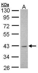
- Submitted by
- Invitrogen Antibodies (provider)
- Main image

- Experimental details
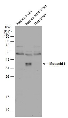
- Western blot analysis of MSI1 was performed by separating 50 µg of various tissue extracts by 12% SDS-PAGE. Proteins were transferred to a membrane and probed with a MSI1 Polyclonal Antibody (Product # PA5-27449) at a dilution of 1:1000. The HRP-conjugated anti-rabbit IgG antibody was used to detect the primary antibody.
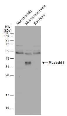
- Submitted by
- Invitrogen Antibodies (provider)
- Main image
- Experimental details
- Western blot analysis of MSI1 was performed by separating 50 µg of various tissue extracts by 12% SDS-PAGE. Proteins were transferred to a membrane and probed with a MSI1 Polyclonal Antibody (Product # PA5-27449) at a dilution of 1:1000. The HRP-conjugated anti-rabbit IgG antibody was used to detect the primary antibody.
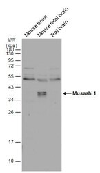
- Submitted by
- Invitrogen Antibodies (provider)
- Main image
- Experimental details
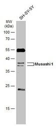
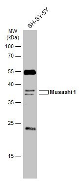
- Western blot analysis of MSI1 was performed by separating 30 µg of whole cell extract by 12% SDS-PAGE. Proteins were transferred to a membrane and probed with a MSI1 Polyclonal Antibody (Product # PA5-27449) at a dilution of 1:1000. The HRP-conjugated anti-rabbit IgG antibody was used to detect the primary antibody.
- Submitted by
- Invitrogen Antibodies (provider)
- Main image
- Experimental details
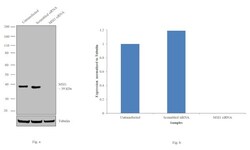
- Knockdown of MSI1 was achieved by transfecting HEK-293 with MSI1 specific siRNAs (Silencer® select Product # s8980; s8979). Western blot analysis (Fig. a) was performed using whole cell extracts from the MSI1 knockdown cells (lane 3), non-specific scrambled siRNA transfected cells (lane 2) and untransfected cells (lane 1). The blots were probed with MSI1 Polyclonal Antibody (Product # PA5-27449, 1:1000 dilution) and Goat anti-Rabbit IgG (H+L) Superclonal™ Secondary Antibody, HRP conjugate (Product # A27036, 0.25µg/mL, 1:4000 dilution). Densitometric analysis of this western blot is shown in histogram (Fig. b). Decrease in signal upon siRNA mediated knock down confirms that antibody is specific to MSI1.

- Submitted by
- Invitrogen Antibodies (provider)
- Main image
- Experimental details
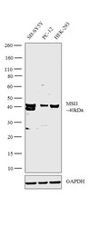
- Western blot analysis was performed on whole cell extracts (30 µg lysate) of SH-SY5Y (Lane 1), PC-12 (Lane 2) and HEK-293 (Lane 3). The blot was probed with Anti-MSI1 Polyclonal Antibody (Product # PA5-27449, 1:2000 dilution) and detected by chemiluminescence Goat anti-Rabbit IgG (H+L) Superclonal™ Secondary Antibody, HRP conjugate (Product # A27036, 0.25 µg/mL, 1:4000 dilution). A 40kDa band corresponding to MSI1 was observed across all the cell lines tested.

Supportive validation
- Submitted by
- Invitrogen Antibodies (provider)
- Main image

- Experimental details
- Immunofluorescent analysis of MSI1 in paraformaldehyde-fixed A549 cells using a MSI1 polyclonal antibody (Product # PA5-27449) at a 1:200 dilution.
- Submitted by
- Invitrogen Antibodies (provider)
- Main image
- Experimental details
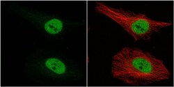
- Immunocytochemistry-Immunofluorescence analysis of MSI1 was performed in HeLa cells fixed in 4% paraformaldehyde at RT for 15 min. Green: MSI1 Polyclonal Antibody (Product # PA5-27449) diluted at 1:500. Red: alpha Tubulin, a cytoskeleton marker.

- Submitted by
- Invitrogen Antibodies (provider)
- Main image
- Experimental details
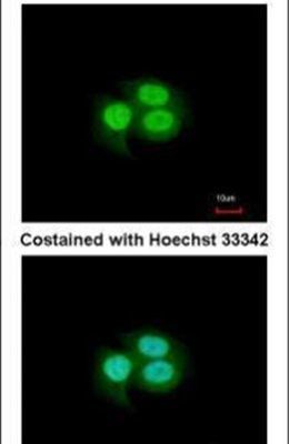
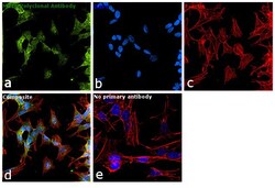
- Immunofluorescence analysis of MSl1 was performed using 70% confluent log phase SH-SY5Y cells. The cells were fixed with 4% paraformaldehyde for 10 minutes, permeabilized with 0.1% Triton™ X-100 for 15 minutes, and blocked with 1% BSA for 1 hour at room temperature. The cells were labeled with anti-MSI1 Polyclonal Antibody (Product # PA5-27449) at 1:100 dilution in 0.1% BSA, incubated at 4 degree Celsius overnight and then labeled with Goat anti-Rabbit IgG (H+L) Superclonal™ Secondary Antibody, Alexa Fluor® 488 conjugate (Product # A27034) at a dilution of 1:2000 for 45 minutes at room temperature (Panel a: green). Nuclei (Panel b: blue) were stained with ProLong™ Diamond Antifade Mountant with DAPI (Product # P36962). F-actin (Panel c: red) was stained with Rhodamine Phalloidin (Product # R415, 1:300). Panel d represents the merged image showing Nuclear and Cytoplasmic localization. Panel e represents control cells with no primary antibody to assess background. The images were captured at 60X magnification

Supportive validation
- Submitted by
- Invitrogen Antibodies (provider)
- Main image
- Experimental details
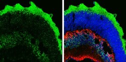
- Immunohistochemistry (Frozen) analysis of MSI1 was performed in frozen sectioned adult mouse retina tissue using MSI1 Polyclonal Antibody (Product # PA5-27449) at a dilution of 1:250 (Green). Red: Protein kinase C alpha staining. Blue: Fluoroshield with DAPI. Antigen Retrieval: EDTA based buffer, pH 8.0 buffer, 15min.
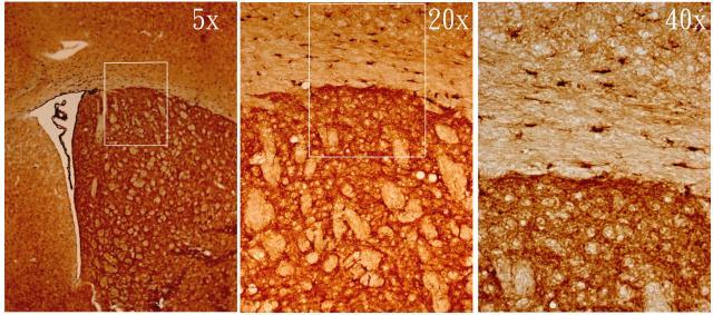
- Submitted by
- Invitrogen Antibodies (provider)
- Main image
- Experimental details
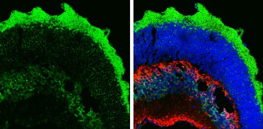
- MSI1 antibody [N3C3] detects MSI1 protein on adult mouse brain by immunohistochemical analysis. Sample: Frozen section of adult mouse brain. MSI1 antibody [N3C3] (Product # PA5-27449) diluted at 1:500.