Explore
 Validate
 Learn
Learn Western blot
Antibody data
- Antibody Data
- Antigen structure
- References [3]
- Comments [0]
- Validations
- Western blot [1]
- Immunocytochemistry [2]
- Immunohistochemistry [1]
Submit
Validation data
Reference
Comment
Report error
- Product number
- AF2628 - Provider product page

- Provider
- R&D Systems
- Product name
- Human/Mouse/Rat Musashi-1 Antibody
- Antibody type
- Polyclonal
- Description
- Antigen Affinity-purified. Detects human Musashi-1 in direct ELISAs and Western blots. In direct ELISAs, less than 1% cross-reactivity with recombinant human Musashi-2 is observed.
- Reactivity
- Human, Mouse, Rat
- Host
- Goat
- Conjugate
- Unconjugated
- Antigen sequence
O43347- Isotype
- IgG
- Vial size
- 100 ug
- Concentration
- LYOPH
- Storage
- Use a manual defrost freezer and avoid repeated freeze-thaw cycles. 12 months from date of receipt, -20 to -70 °C as supplied. 1 month, 2 to 8 °C under sterile conditions after reconstitution. 6 months, -20 to -70 °C under sterile conditions after reconstitution.
Submitted references Rapid generation of OPC-like cells from human pluripotent stem cells for treating spinal cord injury.
Expression and regulation of prostate apoptosis response-4 (Par-4) in human glioma stem cells in drug-induced apoptosis.
Side populations of gastrointestinal cancers are not enriched in stem cells.
Kim DS, Jung SJ, Lee JS, Lim BY, Kim HA, Yoo JE, Kim DW, Leem JW
Experimental & molecular medicine 2017 Jul 28;49(7):e361
Experimental & molecular medicine 2017 Jul 28;49(7):e361
Expression and regulation of prostate apoptosis response-4 (Par-4) in human glioma stem cells in drug-induced apoptosis.
Jagtap JC, Dawood P, Shah RD, Chandrika G, Natesh K, Shiras A, Hegde AS, Ranade D, Shastry P
PloS one 2014;9(2):e88505
PloS one 2014;9(2):e88505
Side populations of gastrointestinal cancers are not enriched in stem cells.
Burkert J, Otto WR, Wright NA
The Journal of pathology 2008 Apr;214(5):564-73
The Journal of pathology 2008 Apr;214(5):564-73
No comments: Submit comment
Supportive validation
- Submitted by
- R&D Systems (provider)
- Main image
- Experimental details
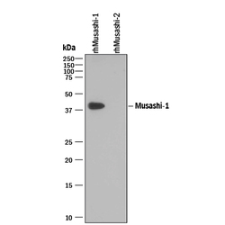
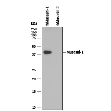
- Detection of Recombinant Human Musashi-1 by Western Blot. Western blot shows 25 ng of Recombinant Human Musashi-1 and Recombinant Human Musashi-2. PVDF Membrane was probed with 0.1 µg/mL of Goat Anti-Human/Mouse/ Rat Musashi-1 Antigen Affinity-purified Polyclonal Antibody (Catalog # AF2628) followed by HRP-conjugated Anti-Goat IgG Secondary Antibody (Catalog # HAF109). A specific band was detected for Musashi-1 at approximately 39 kDa (as indicated). This experiment was conducted under reducing conditions and using Immunoblot Buffer Group 3.
Supportive validation
- Submitted by
- R&D Systems (provider)
- Main image
- Experimental details
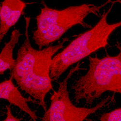
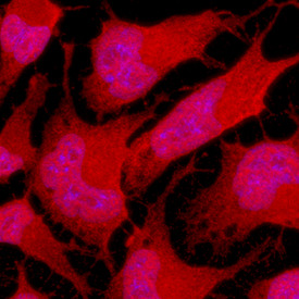
- Musashi-1 in Rat Cortical Stem Cells. Musashi-1 was detected in immersion fixed undifferentiated rat cortical stem cells using Goat Anti-Human/Mouse/Rat Musashi-1 Antigen Affinity-purified Polyclonal Antibody (Catalog # AF2628) at 10 µg/mL for 3 hours at room temperature. Cells were stained using the NorthernLights™ 557-conjugated Anti-Goat IgG Secondary Antibody (red; Catalog # NL001) and counterstained with DAPI (blue). Specific staining was localized to nuclei and cytoplasm. View our protocol for Fluorescent ICC Staining of Stem Cells on Coverslips.
- Submitted by
- R&D Systems (provider)
- Main image
- Experimental details
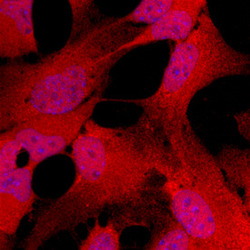
- Musashi-1 in Mouse Cortical Stem Cells. Musashi-1 was detected in immersion fixed undifferentiated mouse cortical stem cells using Goat Anti-Human/Mouse/Rat Musashi-1 Antigen Affinity-purified Polyclonal Antibody (Catalog # AF2628) at 10 µg/mL for 3 hours at room temperature. Cells were stained using the NorthernLights™ 557-conjugated Anti-Goat IgG Secondary Antibody (red; Catalog # NL001) and counter-stained with DAPI (blue). Specific staining was localized to nuclei and cytoplasm. View our protocol for Fluorescent ICC Staining of Stem Cells on Coverslips.

Supportive validation
- Submitted by
- R&D Systems (provider)
- Main image
- Experimental details
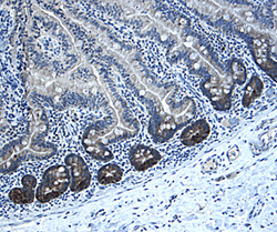
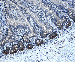
- Musashi-1 in Human Intestine. Musashi-1 was detected in immersion fixed paraffin-embedded sections of human intestine using 1 µg/mL Goat Anti-Human/Mouse/ Rat Musashi-1 Antigen Affinity-purified Polyclonal Antibody (Catalog # AF2628) overnight at 4 °C. Tissue was stained with the Anti-Goat HRP-DAB Cell & Tissue Staining Kit (brown; Catalog # CTS008) and counterstained with hematoxylin (blue). Specific labeling was localized to the cytoplasm of epithelial cells in intestinal glands. View our protocol for Chromogenic IHC Staining of Paraffin-embedded Tissue Sections.