 Explore
 Validate
 Learn
Learn Western blot
 Immunohistochemistry
Antibody data
- Antibody Data
- Antigen structure
- References [1]
- Comments [0]
- Validations
- Immunohistochemistry [1]
Submit
Validation data
Reference
Comment
Report error
- Product number
- AF3486 - Provider product page

- Provider
- Novus Biologicals
- Product name
- Goat Polyclonal Kallikrein 9 Antibody
- Antibody type
- Polyclonal
- Description
- Immunogen affinity purified. Detects human Kallikrein 9 in direct ELISAs and Western blots. In direct ELISAs and Western blots, less than 1% cross-reactivity with recombinant human KLK-1, -3, -4, -5, -6, -7, -8, -10, -11, -12, -13, -14, and -15 is observed.
- Reactivity
- Human
- Host
- Goat
- Isotype
- IgG
- Vial size
- 100 ug
- Concentration
- LYOPH
- Storage
- Use a manual defrost freezer and avoid repeated freeze-thaw cycles. 12 months from date of receipt, -20 to -70 degreesC as supplied. 1 month, 2 to 8 degreesC under sterile conditions after reconstitution. 6 months, -20 to -70 degreesC under sterile conditions after reconstitution.
Submitted references Novel expression of kallikreins, kallikrein-related peptidases and kinin receptors in human pleural mesothelioma.
Chee J, Singh J, Naran A, Misso NL, Thompson PJ, Bhoola KD
Biological chemistry 2007 Nov;388(11):1235-42
Biological chemistry 2007 Nov;388(11):1235-42
No comments: Submit comment
Supportive validation
- Submitted by
- Novus Biologicals (provider)
- Main image
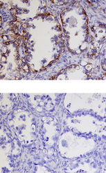
- Experimental details
- Kallikrein 9 in Human Ovarian Cancer Tissue. Kallikrein 9 was detected in immersion fixed paraffin-embedded sections of human ovarian cancer tissue using 15 µg/mL Goat Anti-Human Kallikrein 9 Antigen Affinity-purified Polyclonal Antibody (Catalog # AF3486) overnight at 4 °C. Tissue was stained with the Anti-Goat HRP-DAB Cell & Tissue Staining Kit (brown; Catalog # CTS008) and counterstained with hematoxylin (blue). Lower panel shows a lack of labeling if primary antibodies are omitted and tissue is stained only with secondary antibody followed by incubation with detection reagents. View our protocol for Chromogenic IHC Staining of Paraffin-embedded Tissue Sections.
