Explore
 Validate
 Learn
Learn Western blot
 Immunohistochemistry
Antibody data
- Antibody Data
- Antigen structure
- References [3]
- Comments [0]
- Validations
- Immunohistochemistry [1]
Submit
Validation data
Reference
Comment
Report error
- Product number
- PA3-114 - Provider product page

- Provider
- Invitrogen Antibodies
- Product name
- VPAC2 Polyclonal Antibody
- Antibody type
- Polyclonal
- Antigen
- Synthetic peptide
- Description
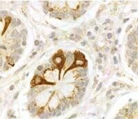
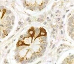
- PA3-114 detects VPAC2 Receptor in human samples. PA3-114 has been successfully used in Western blot, immunocytochemistry and immunohistochemistry procedures. By Western blot, this antibody detects an ~50-65 kDa protein representing VPAC2 Receptor in human samples. In immunohistochemistry, VPAC2 receptor immunoreactivity is predominantly localized to neuroendocrine cells. PA3-114 immunogen is a synthetic peptide, conjugated to KLH, corresponding to the residues L(419) Q F H R G S R A Q S F L Q T E T S V I(438) of human VPAC2 receptor.
- Reactivity
- Human
- Host
- Rabbit
- Isotype
- IgG
- Vial size
- 100 μL
- Concentration
- Conc. Not Determined
- Storage
- -20°C, Avoid Freeze/Thaw Cycles
Submitted references Atypical nuclear localization of VIP receptors in glioma cell lines and patients.
Vasoactive intestinal peptide (VIP) induces malignant transformation of the human prostate epithelial cell line RWPE-1.
Immunocytochemical identification of VPAC1, VPAC2, and PAC1 receptors in normal and neoplastic human tissues with subtype-specific antibodies.
Barbarin A, Séité P, Godet J, Bensalma S, Muller JM, Chadéneau C
Biochemical and biophysical research communications 2014 Nov 28;454(4):524-30
Biochemical and biophysical research communications 2014 Nov 28;454(4):524-30
Vasoactive intestinal peptide (VIP) induces malignant transformation of the human prostate epithelial cell line RWPE-1.
Fernández-Martínez AB, Bajo AM, Isabel Arenas M, Sánchez-Chapado M, Prieto JC, Carmena MJ
Cancer letters 2010 Dec 18;299(1):11-21
Cancer letters 2010 Dec 18;299(1):11-21
Immunocytochemical identification of VPAC1, VPAC2, and PAC1 receptors in normal and neoplastic human tissues with subtype-specific antibodies.
Schulz S, Röcken C, Mawrin C, Weise W, Höllt V, Schulz S
Clinical cancer research : an official journal of the American Association for Cancer Research 2004 Dec 15;10(24):8235-42
Clinical cancer research : an official journal of the American Association for Cancer Research 2004 Dec 15;10(24):8235-42
No comments: Submit comment
Supportive validation
- Submitted by
- Invitrogen Antibodies (provider)
- Main image
- Experimental details
- Immunohistochemical staining of VPAC2 Receptor in human neuroendocrine cells of the small intestine using Product # PA3-114.