 Explore
 Validate
 Learn
LearnAMAb90737
antibody from Atlas Antibodies
Targeting: DICER1
Dicer, HERNA, K12H4.8-LIKE, KIAA0928, MNG1
 Western blot
 Immunocytochemistry
 Immunohistochemistry
Antibody data
- Antibody Data
- Antigen structure
- References [1]
- Comments [0]
- Validations
- Western blot [1]
- Immunocytochemistry [1]
Submit
Validation data
Reference
Comment
Report error
- Product number
- AMAb90737 - Provider product page

- Provider
- Atlas Antibodies
- Proper citation
- Atlas Antibodies Cat#AMAb90737, RRID:AB_2665650
- Product name
- Anti-DICER1
- Antibody type
- Monoclonal
- Description
- Monoclonal Antibody against Human DICER1, Clone ID: CL0378, Gene description: dicer 1, ribonuclease type III, Alternative Gene Names: Dicer, HERNA, K12H4.8-LIKE, KIAA0928, Validated applications: IHC, ICC, WB, Uniprot ID: Q9UPY3, Storage: Store at +4°C for short term storage. Long time storage is recommended at -20°C.
- Reactivity
- Human
- Host
- Mouse
- Conjugate
- Unconjugated
- Isotype
- IgG
- Antibody clone number
- CL0378
- Vial size
- 100 µl
- Concentration
- 0.5 mg/ml
- Storage
- Store at +4°C for short term storage. Long time storage is recommended at -20°C.
- Handling
- The antibody solution should be gently mixed before use.
Submitted references Autophagic degradation of SQSTM1 inhibits ovarian cancer motility by decreasing DICER1 and AGO2 to induce MIRLET7A-3P
Liao C, Ho M, Liang S, Liang C
Autophagy 2018;14(12):2065-2082
Autophagy 2018;14(12):2065-2082
No comments: Submit comment
Enhanced validation
- Submitted by
- Atlas Antibodies (provider)
- Enhanced method
- Genetic validation
- Main image
- Experimental details
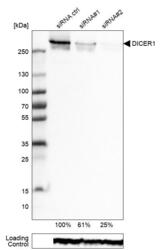
- Western blot analysis in RT-4 cells transfected with control siRNA, target specific siRNA probe #1 and #2, using Anti-DICER1 antibody. Remaining relative intensity is presented. Loading control: Anti-GAPDH.
- Sample type
- Human
- Protocol
- Protocol

Supportive validation
- Submitted by
- Atlas Antibodies (provider)
- Main image
- Experimental details
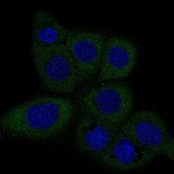
- Immunofluorescence staining of A-431 cells using the anti-DICER1 monoclonal antibody, showing specific staining in the cytosol in green. Microtubule- and nuclear probes are visualized in red and blue, respectively (where available).
- Sample type
- Human
