Explore
 Validate
 Learn
Learn Western blot
 Immunohistochemistry
Antibody data
- Antibody Data
- Antigen structure
- References [2]
- Comments [0]
- Validations
- Western blot [1]
- Other assay [1]
Submit
Validation data
Reference
Comment
Report error
- Product number
- PA1-12565 - Provider product page

- Provider
- Invitrogen Antibodies
- Product name
- CHX10 Polyclonal Antibody
- Antibody type
- Polyclonal
- Antigen
- Other
- Description
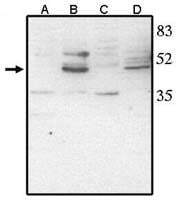
- By Western blot, this antibody detects a ~46 kDa band. A suggested positive control for this product is rat or mouse retinal tissue lysate.
- Reactivity
- Human, Mouse, Rat, Chicken/Avian
- Host
- Sheep
- Isotype
- IgG
- Vial size
- 250 μg
- Concentration
- 1 mg/mL
- Storage
- -20°C, Avoid Freeze/Thaw Cycles
Submitted references Dose-dependent regulation of horizontal cell fate by Onecut family of transcription factors.
Retina Organoid Transplants Develop Photoreceptors and Improve Visual Function in RCS Rats With RPE Dysfunction.
Kreplova M, Kuzelova A, Antosova B, Zilova L, Jägle H, Kozmik Z
PloS one 2020;15(8):e0237403
PloS one 2020;15(8):e0237403
Retina Organoid Transplants Develop Photoreceptors and Improve Visual Function in RCS Rats With RPE Dysfunction.
Lin B, McLelland BT, Aramant RB, Thomas BB, Nistor G, Keirstead HS, Seiler MJ
Investigative ophthalmology & visual science 2020 Sep 1;61(11):34
Investigative ophthalmology & visual science 2020 Sep 1;61(11):34
No comments: Submit comment
Supportive validation
- Submitted by
- Invitrogen Antibodies (provider)
- Main image

- Experimental details
- Western blot analysis of CHX10 using a polyclonal antibody (Product # PA1-12565).
Supportive validation
- Submitted by
- Invitrogen Antibodies (provider)
- Main image
- Experimental details
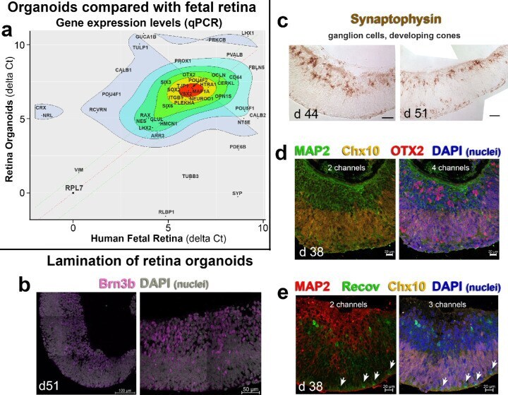
- Figure 1. Characterization of retinal organoids. ( a ) Gene expression scatterplot (mean delta Ct) of d37-d70 retina organoids ( y -axis) versus d105-d145 human fetal retina ( x -axis). Highly co-expressed genes are shown in the center ( red ). The organoids ( n = 17) and fetal tissue ( n = 7) express similar levels of several important developmental genes. Differentially expressed genes are shown in the periphery ( grey ). The retina organoids contain all the major cellular subtypes responsible for support, synaptic integration, and phototransduction. These include glia, ganglion cells, bipolar, horizontal, amacrine, and Muller cells. RPL7 was used as a housekeeping gene. The mean delta Ct values are calculated from biological replicates. ( b ) Confocal microscopy for retinal ganglion cell marker Brn3b ( magenta ) (D51). Nuclei are grey (DAPI). ( c ) Synaptophysin staining of d 44 and d 51 retina organoids, showing mostly developing retinal ganglion cells and cones. ( d ) Confocal microscopy of d38 retinal organoids for MAP 2 ( green ), Chx10 ( gold ), OTX2 ( red ), and DAPI (nuclei, blue ). ( e ) Confocal microscopy of d38 organoids for MAP2 ( red ), Recoverin ( green ), Chx10 ( gold ), and DAPI ( blue ). The w hite arrows point to developing cone photoreceptors. Scale = 100 um ( b ); 50 um ( b , c ); 20 um ( d , e ). DAPI = 4'',6-Diamidino-2-phenylindole.