Explore
 Validate
 Learn
LearnAF793
antibody from Novus Biologicals
Targeting: PPBP
b-TG1, Beta-TG, CTAP3, CTAPIII, CXCL7, LA-PF4, LDGF, MDGF, NAP-2, NAP-2-L1, PBP, SCYB7, TGB, TGB1, THBGB1
 Western blot
 Immunohistochemistry
Antibody data
- Antibody Data
- Antigen structure
- References [2]
- Comments [0]
- Validations
- Immunohistochemistry [1]
- Blocking/Neutralizing [1]
Submit
Validation data
Reference
Comment
Report error
- Product number
- AF793 - Provider product page

- Provider
- Novus Biologicals
- Product name
- Goat Polyclonal CXCL7/Thymus Chemokine-1 Antibody
- Antibody type
- Polyclonal
- Description
- Antigen Affinity-purified. Detects CXCL7/Thymus Chemokine-1 in direct ELISAs and Western blots. In direct ELISAs, approximately 25% cross-reactivity with recombinant rat CXCL7/Thymus Chemokine-1 is observed.
- Reactivity
- Mouse
- Host
- Goat
- Conjugate
- Unconjugated
- Isotype
- IgG
- Vial size
- 100 ug
- Concentration
- LYOPH
- Storage
- Use a manual defrost freezer and avoid repeated freeze-thaw cycles. 12 months from date of receipt, -20 to -70 degreesC as supplied. 1 month, 2 to 8 degreesC under sterile conditions after reconstitution. 6 months, -20 to -70 degreesC under sterile conditions after reconstitution.
Submitted references CXCR4+ CD45- Cells are Niche Forming for Osteoclastogenesis via the SDF-1, CXCL7, and CX3CL1 Signaling Pathways in Bone Marrow.
P2RX7 sensitizes Mac-1/ICAM-1-dependent leukocyte-endothelial adhesion and promotes neurovascular injury during septic encephalopathy.
Goto Y, Aoyama M, Sekiya T, Kakita H, Waguri-Nagaya Y, Miyazawa K, Asai K, Goto S
Stem cells (Dayton, Ohio) 2016 Nov;34(11):2733-2743
Stem cells (Dayton, Ohio) 2016 Nov;34(11):2733-2743
P2RX7 sensitizes Mac-1/ICAM-1-dependent leukocyte-endothelial adhesion and promotes neurovascular injury during septic encephalopathy.
Wang H, Hong LJ, Huang JY, Jiang Q, Tao RR, Tan C, Lu NN, Wang CK, Ahmed MM, Lu YM, Liu ZR, Shi WX, Lai EY, Wilcox CS, Han F
Cell research 2015 Jun;25(6):674-90
Cell research 2015 Jun;25(6):674-90
No comments: Submit comment
Supportive validation
- Submitted by
- Novus Biologicals (provider)
- Main image
- Experimental details
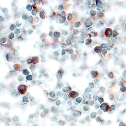
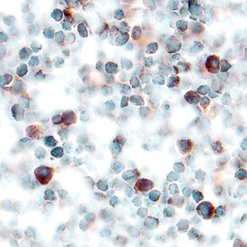
- CXCL7/Thymus Chemokine-1 in Mouse Thymus. CXCL7/Thymus Chemokine-1 was detected in perfusion fixed frozen sections of mouse thymus using Goat Anti-Mouse CXCL7/Thymus Chemokine-1 Antigen Affinity-purified Polyclonal Antibody (Catalog # AF793) at 5 µg/mL overnight at 4 °C. Tissue was stained using the Anti-Goat HRP-DAB Cell & Tissue Staining Kit (brown; Catalog # CTS008) and counterstained with hematoxylin (blue). Specific labeling was localized to the cytoplasm of lymphocytes. View our protocol for Chromogenic IHC Staining of Frozen Tissue Sections.
Supportive validation
- Submitted by
- Novus Biologicals (provider)
- Main image
- Experimental details
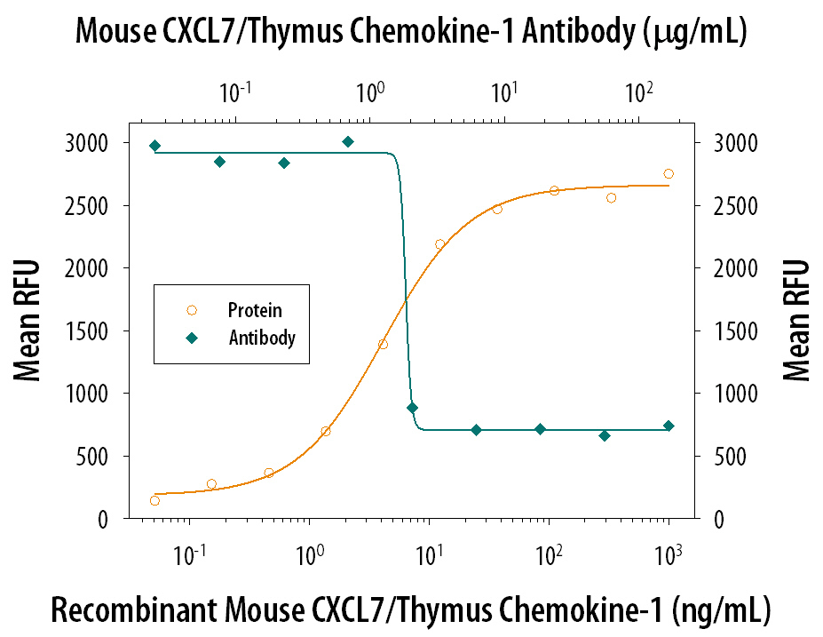
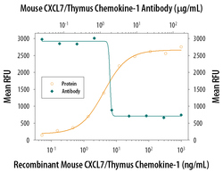
- Chemotaxis Induced by CXCL7/Thymus Chemo-kine-1 and Neutralization by Mouse CXCL7/Thymus Chemo-kine-1 Antibody. Recombinant Mouse CXCL7/Thymus Chemo-kine-1 (Catalog # 1091-CK) chemoattracts the BaF3 mouse pro-B cell line transfected with human CXCR2 in a dose-dependent manner (orange line). The amount of cells that migrated through to the lower chemotaxis chamber was measured by Resazurin (Catalog # AR002). Chemotaxis elicited by Recombinant Mouse CXCL7/Thymus Chemo-kine-1 (40 ng/mL) is neutralized (green line) by increasing concentrations of Goat Anti-Mouse CXCL7/Thymus Chemo-kine-1 Antigen Affinity-purified Polyclonal Antibody (Catalog # AF793). The ND50 is typically 0.8-4 µg/mL.