 Explore
 Validate
 Learn
Learn Immunocytochemistry
Antibody data
- Antibody Data
- Antigen structure
- References [9]
- Comments [0]
- Validations
- Immunocytochemistry [1]
Submit
Validation data
Reference
Comment
Report error
- Product number
- HPA008355 - Provider product page

- Provider
- Atlas Antibodies
- Proper citation
- Atlas Antibodies Cat#HPA008355, RRID:AB_2058277
- Product name
- Anti-ANKS6
- Antibody type
- Polyclonal
- Description
- Polyclonal Antibody against Human ANKS6, Gene description: ankyrin repeat and sterile alpha motif domain containing 6, Alternative Gene Names: ANKRD14, FLJ36928, NPHP16, SAMD6, Validated applications: ICC, Uniprot ID: Q68DC2, Storage: Store at +4°C for short term storage. Long time storage is recommended at -20°C.
- Reactivity
- Human
- Host
- Rabbit
- Conjugate
- Unconjugated
- Isotype
- IgG
- Vial size
- 100 µl
- Concentration
- 0.1 mg/ml
- Storage
- Store at +4°C for short term storage. Long time storage is recommended at -20°C.
- Handling
- The antibody solution should be gently mixed before use.
Submitted references Antagonistic interactions among structured domains in the multivalent Bicc1-ANKS3-ANKS6 protein network govern phase transitioning of target mRNAs
Bicc1 ribonucleoprotein complexes specifying organ laterality are licensed by ANKS6-induced structural remodeling of associated ANKS3
Inactivation of Invs/Nphp2 in renal epithelial cells drives infantile nephronophthisis like phenotypes in mouse
Certain heterozygous variants in the kinase domain of the serine/threonine kinase NEK8 can cause an autosomal dominant form of polycystic kidney disease
Crystal Structure of Bicc1 SAM Polymer and Mapping of Interactions between the Ciliopathy-Associated Proteins Bicc1, ANKS3, and ANKS6
Novel NEK8 Mutations Cause Severe Syndromic Renal Cystic Dysplasia through YAP Dysregulation
The SAM domain of ANKS6 has different interacting partners and mutations can induce different cystic phenotypes
Mutations in ANKS6 Cause a Nephronophthisis-Like Phenotype with ESRD
ANKS6 is a central component of a nephronophthisis module linking NEK8 to INVS and NPHP3
Rothé B, Fortier S, Gagnieux C, Schmuziger C, Constam D
iScience 2023;26(6):106855
iScience 2023;26(6):106855
Bicc1 ribonucleoprotein complexes specifying organ laterality are licensed by ANKS6-induced structural remodeling of associated ANKS3
Lo C, Rothé B, Ikawa Y, Zhang Z, Katoh T, Kajikawa E, Minegishi K, Xiaorei S, Fortier S, Dal Peraro M, Hamada H, Constam D
PLOS Biology 2023;21(9):e3002302
PLOS Biology 2023;21(9):e3002302
Inactivation of Invs/Nphp2 in renal epithelial cells drives infantile nephronophthisis like phenotypes in mouse
Xu W, Li Y, Makova S, Brueckner M, Sun Z
eLife 2023;12
eLife 2023;12
Certain heterozygous variants in the kinase domain of the serine/threonine kinase NEK8 can cause an autosomal dominant form of polycystic kidney disease
Claus L, Chen C, Stallworth J, Turner J, Slaats G, Hawks A, Mabillard H, Senum S, Srikanth S, Flanagan-Steet H, Louie R, Silver J, Lerner-Ellis J, Morel C, Mighton C, Sleutels F, van Slegtenhorst M, van Ham T, Brooks A, Dorresteijn E, Barakat T, Dahan K, Demoulin N, Goffin E, Olinger E, Ambrose J, Arumugam P, Bevers R, Bleda M, Boardman-Pretty F, Boustred C, Brittain H, Caulfield M, Chan G, Elgar G, Fowler T, Giess A, Hamblin A, Henderson S, Hubbard T, Jackson R, Jones L, Kasperaviciute D, Kayikci M, Kousathanas A, Lahnstein L, Leigh S, Leong I, Lopez J, Maleady-Crowe F, McEntagart M, Minneci F, Moutsianas L, Mueller M, Murugaesu N, Need A, O’Donovan P, Odhams C, Patch C, Pereira M, Perez-Gil D, Pullinger J, Rahim T, Rendon A, Rogers T, Savage K, Sawant K, Scott R, Siddiq A, Sieghart A, Smith S, Sosinsky A, Stuckey A, Tanguy M, Taylor Tavares A, Thomas E, Thompson S, Tucci A, Welland M, Williams E, Witkowska K, Wood S, Larsen M, Hertz J, Lilien M, Obeidová L, Seeman T, Stone H, Kerecuk L, Gurgu M, Yousef Yengej F, Ammerlaan C, Rookmaaker M, Hanna C, Rogers R, Duran K, Peters E, Sayer J, van Haaften G, Harris P, Ling K, Mason J, van Eerde A, Steet R
Kidney International 2023;104(5):995-1007
Kidney International 2023;104(5):995-1007
Crystal Structure of Bicc1 SAM Polymer and Mapping of Interactions between the Ciliopathy-Associated Proteins Bicc1, ANKS3, and ANKS6
Rothé B, Leettola C, Leal-Esteban L, Cascio D, Fortier S, Isenschmid M, Bowie J, Constam D
Structure 2018;26(2):209-224.e6
Structure 2018;26(2):209-224.e6
Novel NEK8 Mutations Cause Severe Syndromic Renal Cystic Dysplasia through YAP Dysregulation
Beier D, Grampa V, Delous M, Zaidan M, Odye G, Thomas S, Elkhartoufi N, Filhol E, Niel O, Silbermann F, Lebreton C, Collardeau-Frachon S, Rouvet I, Alessandri J, Devisme L, Dieux-Coeslier A, Cordier M, Capri Y, Khung-Savatovsky S, Sigaudy S, Salomon R, Antignac C, Gubler M, Benmerah A, Terzi F, Attié-Bitach T, Jeanpierre C, Saunier S
PLOS Genetics 2016;12(3):e1005894
PLOS Genetics 2016;12(3):e1005894
The SAM domain of ANKS6 has different interacting partners and mutations can induce different cystic phenotypes
Bakey Z, Bihoreau M, Piedagnel R, Delestré L, Arnould C, de Villiers A, Devuyst O, Hoffmann S, Ronco P, Gauguier D, Lelongt B
Kidney International 2015;88(2):299-310
Kidney International 2015;88(2):299-310
Mutations in ANKS6 Cause a Nephronophthisis-Like Phenotype with ESRD
Taskiran E, Korkmaz E, Gucer S, Kosukcu C, Kaymaz F, Koyunlar C, Bryda E, Chaki M, Lu D, Vadnagara K, Candan C, Topaloglu R, Schaefer F, Attanasio M, Bergmann C, Ozaltin F
Journal of the American Society of Nephrology 2014;25(8):1653-1661
Journal of the American Society of Nephrology 2014;25(8):1653-1661
ANKS6 is a central component of a nephronophthisis module linking NEK8 to INVS and NPHP3
Hoff S, Halbritter J, Epting D, Frank V, Nguyen T, van Reeuwijk J, Boehlke C, Schell C, Yasunaga T, Helmstädter M, Mergen M, Filhol E, Boldt K, Horn N, Ueffing M, Otto E, Eisenberger T, Elting M, van Wijk J, Bockenhauer D, Sebire N, Rittig S, Vyberg M, Ring T, Pohl M, Pape L, Neuhaus T, Elshakhs N, Koon S, Harris P, Grahammer F, Huber T, Kuehn E, Kramer-Zucker A, Bolz H, Roepman R, Saunier S, Walz G, Hildebrandt F, Bergmann C, Lienkamp S
Nature Genetics 2013;45(8):951-956
Nature Genetics 2013;45(8):951-956
No comments: Submit comment
Supportive validation
- Submitted by
- Atlas Antibodies (provider)
- Main image
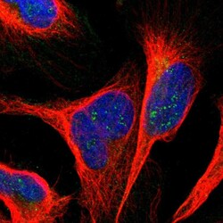
- Experimental details
- Immunofluorescent staining of human cell line U-2 OS shows localization to nucleoli fibrillar center.
- Sample type
- Human
