Explore
 Validate
 Learn
Learn Western blot
Antibody data
- Antibody Data
- Antigen structure
- References [0]
- Comments [0]
- Validations
- Western blot [2]
- Immunocytochemistry [2]
- Immunohistochemistry [1]
- Flow cytometry [1]
Submit
Validation data
Reference
Comment
Report error
- Product number
- APR-008-25UL - Provider product page

- Provider
- Invitrogen Antibodies
- Product name
- P2X7 Receptor (extracellular) Polyclonal Antibody
- Antibody type
- Polyclonal
- Antigen
- Other
- Reactivity
- Human, Mouse, Rat
- Host
- Rabbit
- Isotype
- IgG
- Vial size
- 25 µL
- Concentration
- 0.8 mg/mL
- Storage
- -20° C, Avoid Freeze/Thaw Cycles
No comments: Submit comment
Supportive validation
- Submitted by
- Invitrogen Antibodies (provider)
- Main image
- Experimental details
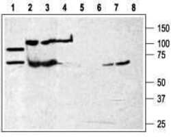
- Western blot analysis of rat brain membranes (lanes 1 and 5) and humanK562 chronic myelogenous leukemia cell line (lanes 2 and 6) andMouseWEHI-231 B cell lymphoma (lanes 3 and 7) and human HL-60 promyelocytic leukemia cell line (lanes 4 and 8): - 1-4. Anti-P2X7 Receptor (extracellular) Antibody (#APR-008), (1:200).5-8. Anti-P2X7 Receptor (extracellular) Antibody ,preincubated with P2X7 Receptor (extracellular) Blocking Peptide (#BLP-PR008).

- Submitted by
- Invitrogen Antibodies (provider)
- Main image

- Experimental details
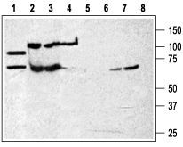
- Western blot analysis of rat brain membranes (lanes 1 and 5) and humanK562 chronic myelogenous leukemia cell line (lanes 2 and 6) andMouseWEHI-231 B cell lymphoma (lanes 3 and 7) and human HL-60 promyelocytic leukemia cell line (lanes 4 and 8): - 1-4. Anti-P2X7 Receptor (extracellular) Antibody (#APR-008), (1:200).5-8. Anti-P2X7 Receptor (extracellular) Antibody ,preincubated with P2X7 Receptor (extracellular) Blocking Peptide (#BLP-PR008).
Supportive validation
- Submitted by
- Invitrogen Antibodies (provider)
- Main image
- Experimental details
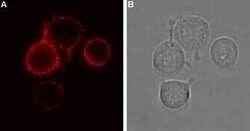
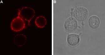
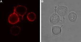
- Expression of P2RX7 in rat RBL cells - Cell surface detection of P2RX7 in intact living rat basophilic leukemia (RBL) cells. A. Extracellular staining of cells using Anti-P2X7 Receptor (extracellular) Antibody (#APR-008), (1:100) followed by goat Anti-rabbit-AlexaFluor-594 secondary Antibody (red). B. Live view of the cells.
- Submitted by
- Invitrogen Antibodies (provider)
- Main image
- Experimental details
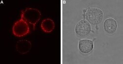
- Expression of P2RX7 in rat RBL cells - Cell surface detection of P2RX7 in intact living rat basophilic leukemia (RBL) cells. A. Extracellular staining of cells using Anti-P2X7 Receptor (extracellular) Antibody (#APR-008), (1:100) followed by goat Anti-rabbit-AlexaFluor-594 secondary Antibody (red). B. Live view of the cells.
Supportive validation
- Submitted by
- Invitrogen Antibodies (provider)
- Main image
- Experimental details
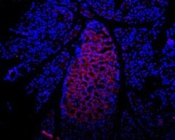
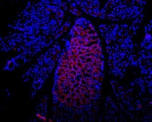
- Expression of P2RX7 in rat pancreas - Immunohistochemical staining of rat pancreas frozen section using Anti-P2X7 Receptor (extracellular) Antibody (#APR-008), (1:100), followed by mouse Anti-rabbit-AlexaFluor-594 secondary Antibody (red). Staining is present in endocrine cells of theIsle of Langerhans. Hoechst 33342 is used as the counterstain.
Supportive validation
- Submitted by
- Invitrogen Antibodies (provider)
- Main image
- Experimental details
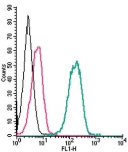
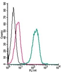
- Cell surface detection of P2X7 Receptor by indirect flow cytometry in live intact human THP-1 monocytic leukemia cells: - (black line) cells. (red) Cells + goat- Anti-rabbit-FITC. (green) Cells + Anti-P2X7 Receptor (extracellular) Antibody (#APR-008), (2.5μg) + goat- Anti-rabbit-FITC.