Explore
 Validate
 Learn
Learn Immunohistochemistry
Antibody data
- Antibody Data
- Antigen structure
- References [3]
- Comments [0]
- Validations
- Immunohistochemistry [3]
- Other assay [2]
Submit
Validation data
Reference
Comment
Report error
- Product number
- PA5-28020 - Provider product page

- Provider
- Invitrogen Antibodies
- Product name
- P2X7 Polyclonal Antibody
- Antibody type
- Polyclonal
- Antigen
- Recombinant full-length protein
- Description
- Predicted reactivity: Mouse (82%), Rat (81%), Dog (91%), Bovine (86%), Guinea pig (83%). Store product as a concentrated solution. Centrifuge briefly prior to opening the vial.
- Reactivity
- Human, Mouse, Rat
- Host
- Rabbit
- Isotype
- IgG
- Vial size
- 100 μL
- Concentration
- 1 mg/mL
- Storage
- Store at 4°C short term. For long term storage, store at -20°C, avoiding freeze/thaw cycles.
Submitted references P2X7 receptor is essential for cross-dressing of bone marrow-derived dendritic cells.
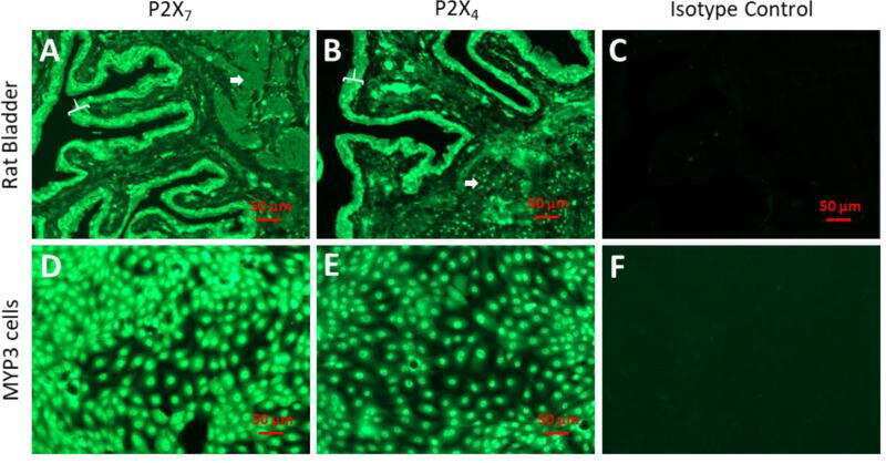
Elevated hydrostatic pressure stimulates ATP release which mediates activation of the NLRP3 inflammasome via P2X(4) in rat urothelial cells.
Targeting of the P2X7 receptor in pancreatic cancer and stellate cells.
Barrera-Avalos C, Briceño P, Valdés D, Imarai M, Leiva-Salcedo E, Rojo LE, Milla LA, Huidobro-Toro JP, Robles-Planells C, Escobar A, Di Virgilio F, Morón G, Sauma D, Acuña-Castillo C
iScience 2021 Dec 17;24(12):103520
iScience 2021 Dec 17;24(12):103520
Elevated hydrostatic pressure stimulates ATP release which mediates activation of the NLRP3 inflammasome via P2X(4) in rat urothelial cells.
Dunton CL, Purves JT, Hughes FM Jr, Jin H, Nagatomi J
International urology and nephrology 2018 Sep;50(9):1607-1617
International urology and nephrology 2018 Sep;50(9):1607-1617
Targeting of the P2X7 receptor in pancreatic cancer and stellate cells.
Giannuzzo A, Saccomano M, Napp J, Ellegaard M, Alves F, Novak I
International journal of cancer 2016 Dec 1;139(11):2540-52
International journal of cancer 2016 Dec 1;139(11):2540-52
No comments: Submit comment
Supportive validation
- Submitted by
- Invitrogen Antibodies (provider)
- Main image
- Experimental details
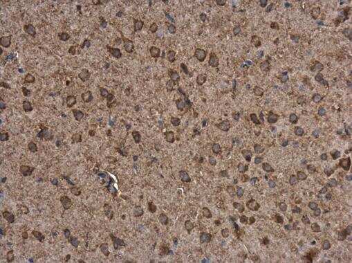
- Immunohistochemistry (Paraffin) analysis of P2X7 was performed in paraffin-embedded rat brain tissue using P2X7 Polyclonal Antibody (Product # PA5-28020) at a dilution of 1:500.
- Submitted by
- Invitrogen Antibodies (provider)
- Main image
- Experimental details
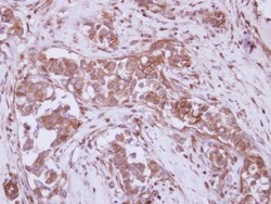
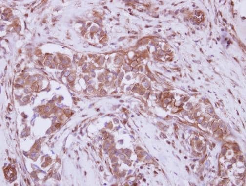
- Immunohistochemical analysis of paraffin-embedded human breast cancer, using P2X7 (Product # PA5-28020) antibody at 1:250 dilution. Antigen Retrieval: Citrate buffer, pH 6.0, 15 min.
- Submitted by
- Invitrogen Antibodies (provider)
- Main image

- Experimental details
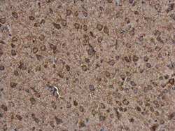
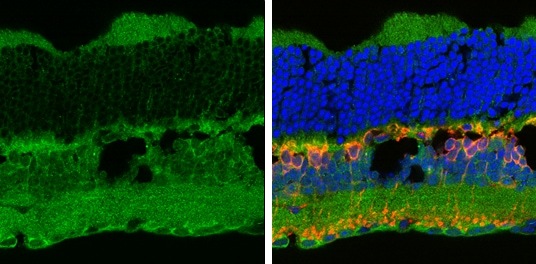
- Immunohistochemistry (Frozen) analysis of P2X7 was performed in frozen sectioned adult mouse retina tissue using P2X7 Polyclonal Antibody (Product # PA5-28020) at a dilution of 1:250 (Green). Red: Protein kinase C alpha staining. Blue: Fluoroshield with DAPI. Antigen Retrieval: Citrate buffer, pH 6.0, 15 min.
Supportive validation
- Submitted by
- Invitrogen Antibodies (provider)
- Main image
- Experimental details
- NULL
- Submitted by
- Invitrogen Antibodies (provider)
- Main image
- Experimental details
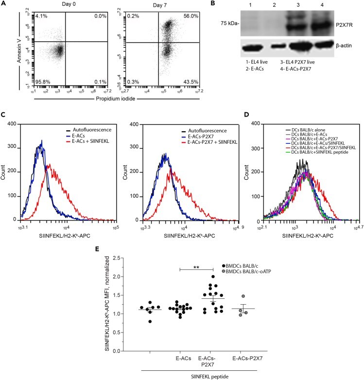
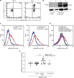
- Figure 3 CD8+ OT-1 lymphocytes are activated by P2X7-dependent cross-dressing (A) ACs generated from EL4 cells (E-ACs) by nutrient deprivation-induced cell death were identified with Annexin V (AV) and Propidium Iodide (PI) on the seventh day of culture. Representative dot plots of flow cytometry analysis show two populations of E-ACs: late Apoptotic (PI + AV+) and early apoptotic (PI-AV+) E-ACs. Data represents one of three independent experiments. Only PI-AV- cells are present at the beginning of treatment. (B) Western blot for P2X7 receptor from EL4 live cells, E-ACs, EL4 P2X7 live cells, and E-ACs-P2X7, 1-4 lanes, left to right, respectively. The 75 kDa band corresponds to the molecular weight of the P2X7 receptor. (C) The SIINFEKL/H2-K b presence was evaluated by flow cytometry in ACs generated from both parental (E-ACs, left side) and P2X7 overexpressing (E-ACs-P2X7, right side) H-2K b EL4 cells. SIINFEKL/H2-K b in unloaded ACs (blue) and peptide-loaded ACs (red) were compared against basal fluorescence (no antibody, black). (D) Evaluation of antigen/MHC (SIINFEKL/H2-K b complex) transference from ACs to the surface of allogenic (H-2 d ) BMDCs-BALB/c. A representative histogram from 15 independent experiments of the SIINFEKL/H2-K b detected in CD11+ BM-DCs challenged with E-ACs (gray), E-ACs-P2X7 (pink), E-ACs SIINFEKL (blue), and E-ACs-P2X7/SIINFEKL (red). As controls, BMDCs-BALB/c not challenged (black) and challenged with SIINFEKL peptide (green) were included. (E) T