Explore
 Validate
 Learn
Learn Western blot
Antibody data
- Antibody Data
- Antigen structure
- References [1]
- Comments [0]
- Validations
- Western blot [2]
- Immunocytochemistry [1]
- Immunohistochemistry [1]
Submit
Validation data
Reference
Comment
Report error
- Product number
- GTX109663 - Provider product page

- Provider
- GeneTex
- Proper citation
- GeneTex Cat#GTX109663, RRID:AB_1949956
- Product name
- CDT1 antibody
- Antibody type
- Polyclonal
- Reactivity
- Human
- Host
- Rabbit
Submitted references Cell cycle kinetic analysis of colorectal neoplasms using a new automated immunohistochemistry-based cell cycle detection method.
Tomono A, Itoh T, Yanagita E, Imagawa N, Kakeji Y
Medicine 2015 Jan;94(4):e501
Medicine 2015 Jan;94(4):e501
No comments: Submit comment
Supportive validation
- Submitted by
- GeneTex (provider)
- Main image
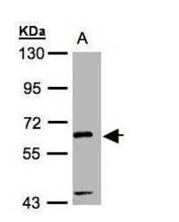
- Experimental details
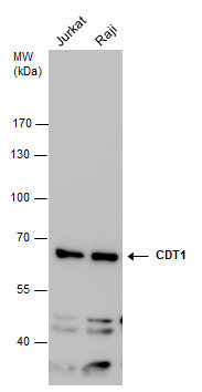
- Sample(30 ug whole cell lysate)A:Raji (GTX27908)10% SDS PAGEGTX109663 diluted at 1:1000
- Validation comment
- WB
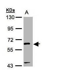
- Submitted by
- GeneTex (provider)
- Main image
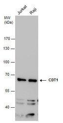
- Experimental details
- CDT1 antibody detects CDT1 protein by western blot analysis. Various whole cell extracts (30 £gg) were separated by 7.5% SDS-PAGE, and the membrane was blotted with CDT1 antibody (GTX109663) diluted at a dilution of 1:1000.
Supportive validation
- Submitted by
- GeneTex (provider)
- Main image
- Experimental details
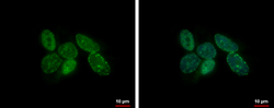
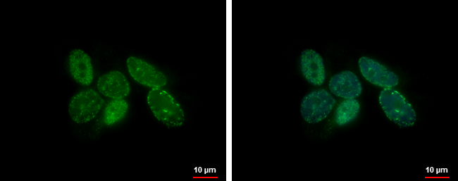
- CDT1 antibody detects CDT1 protein at nucleus by immunofluorescent analysis.Sample: SK-N-AS cells were fixed in 4% paraformaldehyde at RT for 15 min.Green: CDT1 protein stained by CDT1 antibody (GTX109663) diluted at 1:500.Blue: Hoechst 33342 staining.
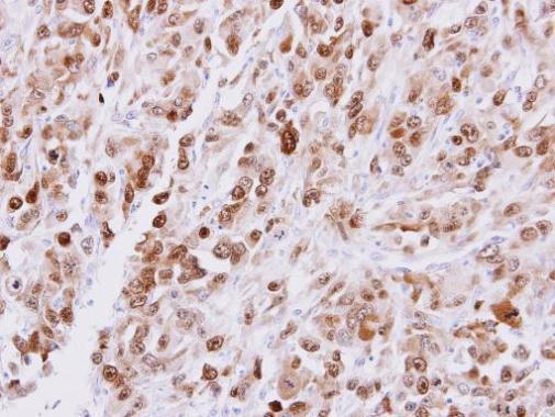
Supportive validation
- Submitted by
- GeneTex (provider)
- Main image
- Experimental details
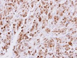
- Immunohistochemical analysis of paraffin-embedded ES2 xenograft, using CDT1(GTX109663) antibody at 1:500 dilution.