Explore
 Validate
 Learn
Learn Western blot
 ELISA
ELISAAntibody data
- Antibody Data
- Antigen structure
- References [0]
- Comments [0]
- Validations
- Western blot [1]
Submit
Validation data
Reference
Comment
Report error
- Product number
- A01127 - Provider product page

- Provider
- Boster Biological Technology
- Product name
- Anti-14-3-3 sigma/SFN Antibody Picoband™
- Antibody type
- Polyclonal
- Description
- Polyclonal antibody for STRATIFIN/SFN detection. Host: Rabbit.Size: 100μg/vial. Tested applications: ELISA. Reactive species: Human. STRATIFIN/SFN information: Subcellular Localization: Cytoplasm. Nucleus . Secreted. May be secreted by a non-classical secretory pathway; Tissue Specificity: Present mainly in tissues enriched in stratified squamous keratinizing epithelium.
- Reactivity
- Human, Mouse, Rat
- Host
- Rabbit
- Vial size
- 100μg/vial
- Concentration
- 0.5-1mg/ml, actual concentration vary by lot. Use suggested dilution ratio to decide dilution procedure.
- Storage
- At -20°C for one year. After reconstitution, at 4°C for one month. It can also be aliquoted and stored frozen at -20°C for a longer time. Avoid repeated freezing and thawing.
- Handling
- Add 0.2ml of distilled water will yield a concentration of 500ug/ml.
No comments: Submit comment
Supportive validation
- Submitted by
- Boster Biological Technology (provider)
- Main image
- Experimental details
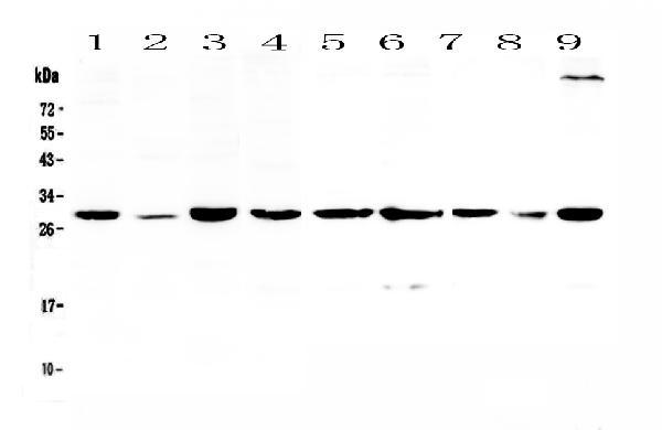
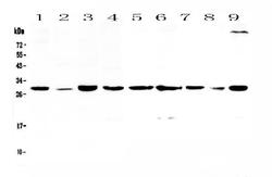
- Western blot analysis of 14-3-3 sigma using anti-14-3-3 sigma antibody (A01127). Electrophoresis was performed on a 5-20% SDS-PAGE gel at 70V (Stacking gel) / 90V (Resolving gel) for 2-3 hours. The sample well of each lane was loaded with 50ug of sample under reducing conditions. Lane 1: human Hela cell lysate,Lane 2: human placenta tissue lysate,Lane 3: human MCF-7 cell lysate,Lane 4: human 22RV1 cell lysate,Lane 5: rat lung tissue lysate,Lane 6: rat PC-12 cell lysate,Lane 7: mouse lung tissue lysate,Lane 8: mouse HEPA1-6 cell lysate,Lane 9: mouse NIH3T3 cell lysate. After Electrophoresis, proteins were transferred to a Nitrocellulose membrane at 150mA for 50-90 minutes. Blocked the membrane with 5% Non-fat Milk/ TBS for 1.5 hour at RT. The membrane was incubated with rabbit anti-14-3-3 sigma antigen affinity purified polyclonal antibody (Catalog # A01127) at 0.5 μg/mL overnight at 4°C, then washed with TBS-0.1%Tween 3 times with 5 minutes each and probed with a goat anti-rabbit IgG-HRP secondary antibody at a dilution of 1:10000 for 1.5 hour at RT. The signal is developed using an Enhanced Chemiluminescent detection (ECL) kit (Catalog # EK1002) with Tanon 5200 system. A specific band was detected for 14-3-3 sigma at approximately 28KD. The expected band size for 14-3-3 sigma is at 28KD.
- Additional image