Explore
 Validate
 Learn
Learn Western blot
Antibody data
- Antibody Data
- Antigen structure
- References [0]
- Comments [0]
- Validations
- Western blot [6]
- Immunocytochemistry [1]
- Immunohistochemistry [2]
Submit
Validation data
Reference
Comment
Report error
- Product number
- PA5-27147 - Provider product page

- Provider
- Invitrogen Antibodies
- Product name
- 14-3-3 sigma Polyclonal Antibody
- Antibody type
- Polyclonal
- Antigen
- Synthetic peptide
- Description
- Recommended positive controls: 293T, A431, HeLa, HepG2, Neuro2A, GL261, PC-12, A549.
- Concentration
- 1 mg/mL
No comments: Submit comment
Supportive validation
- Submitted by
- Invitrogen Antibodies (provider)
- Main image

- Experimental details
- Western blot analysis of 14-3-3 sigma using A) 30 µg Neuro2A whole cell lysate and B) 30 µg GL261 whole cell lysate. Samples were loaded onto a 12% SDS-PAGE gel and probed with a 14-3-3 sigma polyclonal antibody (Product # PA5-27147) at a dilution of 1:3000.

- Submitted by
- Invitrogen Antibodies (provider)
- Main image

- Experimental details
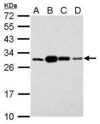
- 14-3-3 sigma Polyclonal Antibody detects SFN protein by western blot analysis. A. 30 µg 293T whole cell lysate/extract. B. 30 µg A431 whole cell lysate/extract. C. 30 µg HeLa whole cell lysate/extract. D. 30 µg HepG2 whole cell lysate/extract.12% SDS-PAGE.14-3-3 sigma Polyclonal Antibody (Product # PA5-27147) dilution: 1:3,000. The HRP-conjugated anti-rabbit IgG antibody was used to detect the primary antibody.

- Submitted by
- Invitrogen Antibodies (provider)
- Main image

- Experimental details
- Western blot analysis of 14-3-3 sigma was performed by separating 30 µg of whole cell extract by 12% SDS-PAGE. Proteins were transferred to a membrane and probed with a 14-3-3 sigma Polyclonal Antibody (Product # PA5-27147) at a dilution of 1:3000. The HRP-conjugated anti-rabbit IgG antibody was used to detect the primary antibody.

- Submitted by
- Invitrogen Antibodies (provider)
- Main image
- Experimental details
- 14-3-3 sigma Polyclonal Antibody detects SFN protein by western blot analysis. A. 30 µg 293T whole cell lysate/extract. B. 30 µg A431 whole cell lysate/extract. C. 30 µg HeLa whole cell lysate/extract. D. 30 µg HepG2 whole cell lysate/extract.12% SDS-PAGE.14-3-3 sigma Polyclonal Antibody (Product # PA5-27147) dilution: 1:3,000. The HRP-conjugated anti-rabbit IgG antibody was used to detect the primary antibody.

- Submitted by
- Invitrogen Antibodies (provider)
- Main image

- Experimental details
- 14-3-3 sigma Polyclonal Antibody detects SFN protein by western blot analysis. A. 30 µg Neuro2A whole cell lysate/extract. B. 30 µg GL261 whole cell lysate/extract.12% SDS-PAGE.14-3-3 sigma Polyclonal Antibody (Product # PA5-27147) dilution: 1:3,000. The HRP-conjugated anti-rabbit IgG antibody was used to detect the primary antibody.

- Submitted by
- Invitrogen Antibodies (provider)
- Main image

- Experimental details
- 14-3-3 sigma Polyclonal Antibody detects SFN protein by western blot analysis. A. 30 µg PC-12 whole cell lysate/extract.12% SDS-PAGE.14-3-3 sigma Polyclonal Antibody (Product # PA5-27147) dilution: 1:3,000. The HRP-conjugated anti-rabbit IgG antibody was used to detect the primary antibody.

Supportive validation
- Submitted by
- Invitrogen Antibodies (provider)
- Main image

- Experimental details
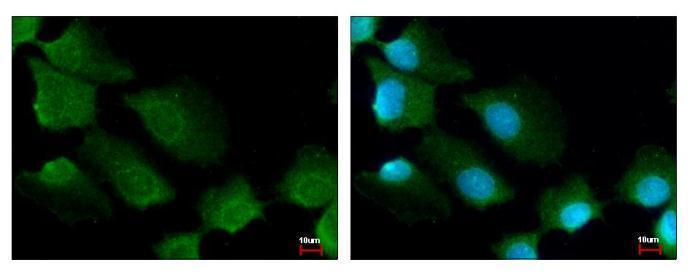
- 14-3-3 sigma Polyclonal Antibody [C1C3] detects 14-3-3 sigma protein at cytoplasm by immunofluorescent analysis. Sample: A431 cells were fixed in ice-cold Methanol for 5 min. Green: 14-3-3 sigma protein stained by 14-3-3 sigma Polyclonal Antibody [C1C3] (Product # PA5-27147) diluted at 1:500. Blue: Hoechst 33343 staining.
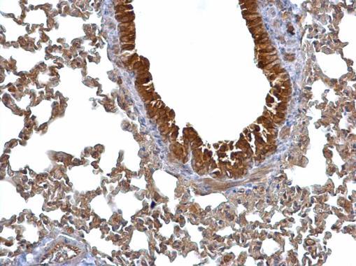
Supportive validation
- Submitted by
- Invitrogen Antibodies (provider)
- Main image
- Experimental details
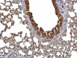
- 14-3-3 sigma Polyclonal Antibody detects 14-3-3 sigma protein at cytosol on mouse lung by immunohistochemical analysis. Sample: Paraffin-embedded mouse lung.14-3-3 sigma Polyclonal Antibody (Product # PA5-27147) dilution: 1:500. Antigen Retrieval: EDTA based buffer, pH 8.0, 15 min.
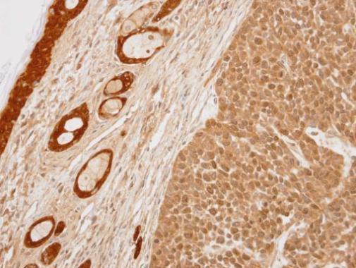
- Submitted by
- Invitrogen Antibodies (provider)
- Main image
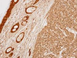
- Experimental details
- Immunohistochemical analysis of paraffin-embedded 59T xenograft, using 14-3-3 sigma (Product # PA5-27147) antibody at 1:100 dilution. Antigen Retrieval: EDTA based buffer, pH 8.0, 15 min.