Explore
 Validate
 Learn
Learn Western blot
 Immunocytochemistry
Antibody data
- Antibody Data
- Antigen structure
- References [2]
- Comments [0]
- Validations
- Western blot [1]
Submit
Validation data
Reference
Comment
Report error
- Product number
- PA1797 - Provider product page

- Provider
- Boster Biological Technology
- Product name
- Anti-14-3-3 sigma/SFN Antibody
- Antibody type
- Polyclonal
- Description
- Polyclonal antibody for STRATIFIN/SFN detection. Host: Rabbit.Size: 100μg/vial. Tested applications: WB, IHC-P, ICC, ICC/IF, FCM. Reactive species: Human;Mouse;Rat. STRATIFIN/SFN information: Molecular Weight: 27774 MW; Subcellular Localization: Cytoplasm. Nucleus . Secreted. May be secreted by a non-classical secretory pathway; Tissue Specificity: Present mainly in tissues enriched in stratified squamous keratinizing epithelium.
- Reactivity
- Human, Mouse, Rat
- Host
- Rabbit
- Vial size
- 100μg/vial
- Concentration
- Add 0.2ml of distilled water will yield a concentration of 500ug/ml.
- Storage
- At -20°C for one year. After reconstitution, at 4°C for one month. It can also be aliquoted and stored frozen at -20°C for a longer time. Avoid repeated freezing and thawing.
- Handling
- Add 0.2ml of distilled water will yield a concentration of 500ug/ml.
Submitted references Protective Effect of Dictyophora Polysaccharides on Sodium Arsenite-Induced Hepatotoxicity: A Proteomics Study.
Histone acetyltransferase inhibitor II induces apoptosis in glioma cell lines via the p53 signaling pathway.
Hu T, Shen L, Huang Q, Wu C, Zhang H, Zeng Q, Wang G, Wei S, Zhang S, Zhang J, Khan NU, Shen X, Luo P
Frontiers in pharmacology 2021;12:749035
Frontiers in pharmacology 2021;12:749035
Histone acetyltransferase inhibitor II induces apoptosis in glioma cell lines via the p53 signaling pathway.
Xu LX, Li ZH, Tao YF, Li RH, Fang F, Zhao H, Li G, Li YH, Wang J, Feng X, Pan J
Journal of experimental & clinical cancer research : CR 2014 Dec 19;33(1):108
Journal of experimental & clinical cancer research : CR 2014 Dec 19;33(1):108
No comments: Submit comment
Supportive validation
- Submitted by
- Boster Biological Technology (provider)
- Main image
- Experimental details
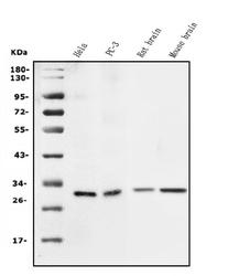
- Western blot analysis of SFN using anti-SFN antibody (PA1797). Electrophoresis was performed on a 5-20% SDS-PAGE gel at 70V (Stacking gel) / 90V (Resolving gel) for 2-3 hours. The sample well of each lane was loaded with 50ug of sample under reducing conditions. Lane 1: human Hela whole cell lysates, Lane 2: human PC-3 whole cell lysates, Lane 3: rat brain tissue lysates, Lane 4: mouse brain tissue lysates. After Electrophoresis, proteins were transferred to a Nitrocellulose membrane at 150mA for 50-90 minutes. Blocked the membrane with 5% Non-fat Milk/ TBS for 1.5 hour at RT. The membrane was incubated with rabbit anti-SFN antigen affinity purified polyclonal antibody (Catalog # PA1797) at 0.5 μg/mL overnight at 4°C, then washed with TBS-0.1%Tween 3 times with 5 minutes each and probed with a goat anti-rabbit IgG-HRP secondary antibody at a dilution of 1:10000 for 1.5 hour at RT. The signal is developed using an Enhanced Chemiluminescent detection (ECL) kit (Catalog # EK1002) with Tanon 5200 system. A specific band was detected for SFN at approximately 28KD. The expected band size for SFN is at 28KD.
- Additional image