 Explore
 Validate
 Learn
Learn Western blot
 ELISA
ELISAAntibody data
- Antibody Data
- Antigen structure
- References [3]
- Comments [0]
- Validations
- Western blot [1]
- Flow cytometry [1]
Submit
Validation data
Reference
Comment
Report error
- Product number
- MAB1180-100 - Provider product page

- Provider
- R&D Systems
- Product name
- Human DPPIV/CD26 Antibody
- Antibody type
- Monoclonal
- Description
- Protein A or G purified from hybridoma culture supernatant. Detects human DPPIV/CD26 in ELISAs and Western blots. In ELISAs and Western blots, no cross-reactivity with recombinant human Cathepsin A or recombinant mouse DPPIV is observed.
- Reactivity
- Human
- Host
- Rat
- Conjugate
- Unconjugated
- Antigen sequence
Q53TN1- Isotype
- IgG
- Antibody clone number
- 222113
- Vial size
- 100 ug
- Concentration
- LYOPH
- Storage
- Use a manual defrost freezer and avoid repeated freeze-thaw cycles. 12 months from date of receipt, -20 to -70 °C as supplied. 1 month, 2 to 8 °C under sterile conditions after reconstitution. 6 months, -20 to -70 °C under sterile conditions after reconstitution.
Submitted references Engineering a stable CHO cell line for the expression of a MERS-coronavirus vaccine antigen.
Experimental infection of dromedaries with Middle East respiratory syndrome-Coronavirus is accompanied by massive ciliary loss and depletion of the cell surface receptor dipeptidyl peptidase 4.
Involvement of DPP-IV catalytic residues in enzyme-saxagliptin complex formation.
Nyon MP, Du L, Tseng CK, Seid CA, Pollet J, Naceanceno KS, Agrawal A, Algaissi A, Peng BH, Tai W, Jiang S, Bottazzi ME, Strych U, Hotez PJ
Vaccine 2018 Mar 27;36(14):1853-1862
Vaccine 2018 Mar 27;36(14):1853-1862
Experimental infection of dromedaries with Middle East respiratory syndrome-Coronavirus is accompanied by massive ciliary loss and depletion of the cell surface receptor dipeptidyl peptidase 4.
Haverkamp AK, Lehmbecker A, Spitzbarth I, Widagdo W, Haagmans BL, Segalés J, Vergara-Alert J, Bensaid A, van den Brand JMA, Osterhaus ADME, Baumgärtner W
Scientific reports 2018 Jun 27;8(1):9778
Scientific reports 2018 Jun 27;8(1):9778
Involvement of DPP-IV catalytic residues in enzyme-saxagliptin complex formation.
Metzler WJ, Yanchunas J, Weigelt C, Kish K, Klei HE, Xie D, Zhang Y, Corbett M, Tamura JK, He B, Hamann LG, Kirby MS, Marcinkeviciene J
Protein science : a publication of the Protein Society 2008 Feb;17(2):240-50
Protein science : a publication of the Protein Society 2008 Feb;17(2):240-50
No comments: Submit comment
Supportive validation
- Submitted by
- R&D Systems (provider)
- Main image

- Experimental details
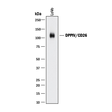
- Detection of Human DPPIV/CD26 by Western Blot. Western blot shows lysate of LoVo human colorectal adenocarcinoma cell line. PVDF membrane was probed with 2 µg/mL of Rat Anti-Human DPPIV/CD26 Monoclonal Antibody (Catalog # MAB1180) followed by HRP-conjugated Anti-Rat IgG Secondary Antibody (Catalog # HAF005). A specific band was detected for DPPIV/CD26 at approximately 110 kDa (as indicated). This experiment was conducted under reducing conditions and using Immunoblot Buffer Group 1.
Supportive validation
- Submitted by
- R&D Systems (provider)
- Main image
- Experimental details
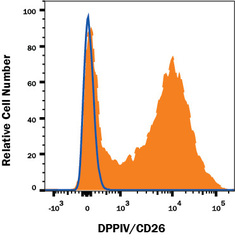
- Detection of DPPIV/CD26 in Human Blood Lymphocytes by Flow Cytometry. Human peripheral blood lymphocytes were stained with Rat Anti-Human DPPIV/CD26 Monoclonal Antibody (Catalog # MAB1180, filled histogram) or isotype control antibody (Catalog # MAB006, open histogram) followed by Anti-mouse PE-conjugated secondary antibody (Catalog # F0102B). View our protocol for Staining Membrane-associated Proteins.
