 Explore
 Validate
 Learn
Learn Western blot
 Immunocytochemistry
Antibody data
- Antibody Data
- Antigen structure
- References [0]
- Comments [0]
- Validations
- Western blot [1]
- Immunohistochemistry [1]
Submit
Validation data
Reference
Comment
Report error
- Product number
- NBP1-92689 - Provider product page

- Provider
- Novus Biologicals
- Proper citation
- Novus Cat#NBP1-92689, RRID:AB_11031596
- Product name
- Mouse Monoclonal Alpha Fodrin Antibody
- Antibody type
- Monoclonal
- Description
- Immunogen affinity purified.
- Reactivity
- Human, Mouse, Rat
- Host
- Mouse
- Isotype
- IgG
- Vial size
- 0.1 ml
- Concentration
- 1 mg/ml
- Storage
- Store at 4C short term. Aliquot and store at -20C long term. Avoid freeze-thaw cycles.
No comments: Submit comment
Supportive validation
- Submitted by
- Novus Biologicals (provider)
- Main image

- Experimental details
- Western Blot: Alpha Fodrin Antibody (3D7) [NBP1-92689] - Analysis of neural tissue and cell lysates using mouse mAb to alpha-II spectrin (NBP1-92689, green). [1] protein standard, [2] rat whole brain, [3] rat spinal cord, [4] mouse whole brain, [5] mouse spinal cord, [6] NIH-3T3, [7] HEK293, [8] HeLa, [9] SH-SY5Y, [10] C6 cells. A prominent band at about 250-260 kDa represents the intact alpha-II spectrin heavy chain in neural tissues or cell lines, but not in HeLa.

Supportive validation
- Submitted by
- Novus Biologicals (provider)
- Main image
- Experimental details
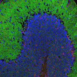
- Immunohistochemistry: Alpha Fodrin Antibody (3D7) [NBP1-92689] - Immunofluorescent image of rat cerebellum stained with NBP1-92689 antibody to alpha-II spectrin, dilution 1:2,000, in green, chicken polyclonal antibody to GFAP, dilution 1:5,000, in red. The spectrin antibody stains the submembraneous cytoskeleton on neurons and strongly reveals the cell bodies and dendrites of Purkinje cells, while the GFAP antibody stains the processes of Bergmann glia and astrocytes.
