Explore
 Validate
 Learn
Learn Western blot
 Immunocytochemistry
Antibody data
- Antibody Data
- Antigen structure
- References [4]
- Comments [0]
- Validations
- Immunocytochemistry [1]
- Immunohistochemistry [1]
Submit
Validation data
Reference
Comment
Report error
- Product number
- HPA006360 - Provider product page

- Provider
- Atlas Antibodies
- Proper citation
- Atlas Antibodies Cat#HPA006360, RRID:AB_1079082
- Product name
- Anti-HPRT1
- Antibody type
- Polyclonal
- Description
- Polyclonal Antibody against Human HPRT1, Gene description: hypoxanthine phosphoribosyltransferase 1, Alternative Gene Names: HGPRT, HPRT, Validated applications: ICC, WB, IHC, Uniprot ID: P00492, Storage: Store at +4°C for short term storage. Long time storage is recommended at -20°C.
- Reactivity
- Human
- Host
- Rabbit
- Conjugate
- Unconjugated
- Isotype
- IgG
- Vial size
- 100 µl
- Concentration
- 0.2 mg/ml
- Storage
- Store at +4°C for short term storage. Long time storage is recommended at -20°C.
- Handling
- The antibody solution should be gently mixed before use.
Submitted references Regulating PCCA gene expression by modulation of pseudoexon splicing patterns to rescue enzyme activity in propionic acidemia
DeepCLIP: predicting the effect of mutations on protein–RNA binding with deep learning
Blocking of an intronic splicing silencer completely rescues IKBKAP exon 20 splicing in familial dysautonomia patient cells
Global identification of hnRNP A1 binding sites for SSO-based splicing modulation
Spangsberg Petersen U, Dembic M, Martínez-Pizarro A, Richard E, Holm L, Havelund J, Doktor T, Larsen M, Færgeman N, Desviat L, Andresen B
Molecular Therapy - Nucleic Acids 2024;35(1):102101
Molecular Therapy - Nucleic Acids 2024;35(1):102101
DeepCLIP: predicting the effect of mutations on protein–RNA binding with deep learning
Andresen B, Baumbach J, Hartung A, Hansen M, Bruun G, Holm L, Petersen U, Larsen S, Doktor T, Grønning A
Nucleic Acids Research 2020
Nucleic Acids Research 2020
Blocking of an intronic splicing silencer completely rescues IKBKAP exon 20 splicing in familial dysautonomia patient cells
Bruun G, Bang J, Christensen L, Brøner S, Petersen U, Guerra B, Grønning A, Doktor T, Andresen B
Nucleic Acids Research 2018;46(15):7938-7952
Nucleic Acids Research 2018;46(15):7938-7952
Global identification of hnRNP A1 binding sites for SSO-based splicing modulation
Bruun G, Doktor T, Borch-Jensen J, Masuda A, Krainer A, Ohno K, Andresen B
BMC Biology 2016;14(1)
BMC Biology 2016;14(1)
No comments: Submit comment
Supportive validation
- Submitted by
- Atlas Antibodies (provider)
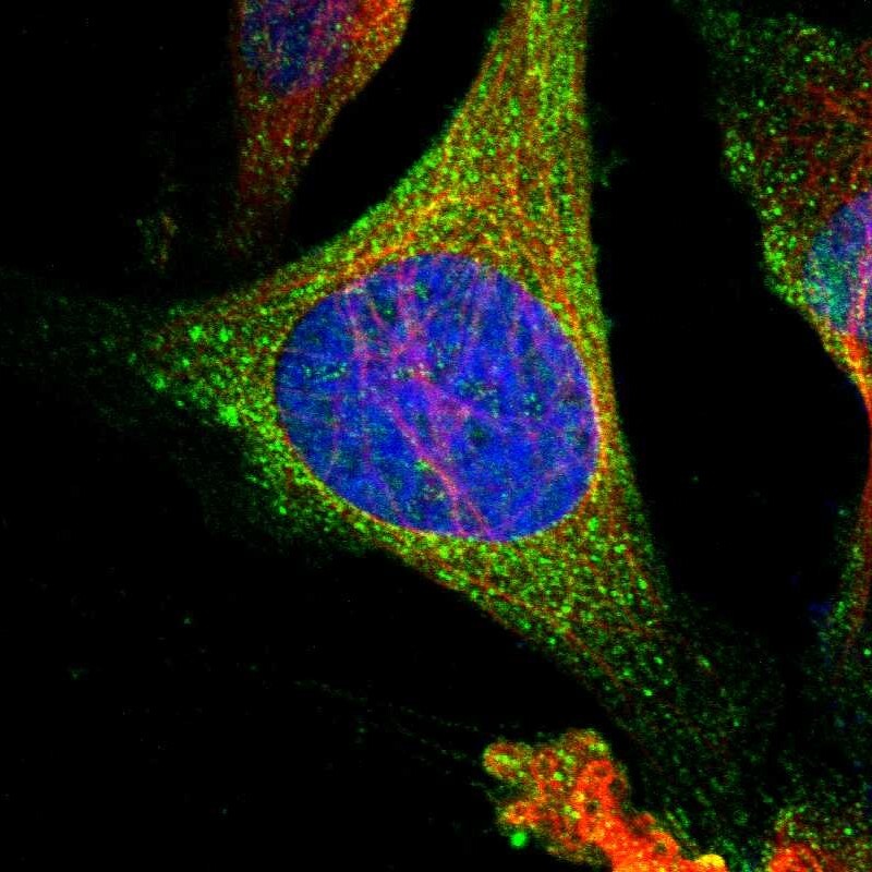
- Main image

- Experimental details
- Immunofluorescent staining of human cell line U-2 OS shows localization to cytosol.
- Sample type
- Human
Supportive validation
- Submitted by
- Atlas Antibodies (provider)
- Enhanced method
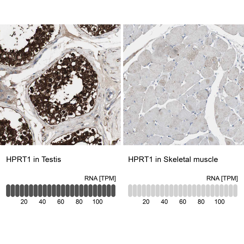
- Orthogonal validation
- Main image

- Experimental details
- Immunohistochemistry analysis in human testis and skeletal muscle tissues using HPA006360 antibody. Corresponding HPRT1 RNA-seq data are presented for the same tissues.
- Sample type
- Human
- Protocol
- Protocol