 Explore
 Validate
 Learn
Learn Western blot
 ELISA
ELISA Flow cytometry
Antibody data
- Antibody Data
- Antigen structure
- References [1]
- Comments [0]
- Validations
- Western blot [5]
Submit
Validation data
Reference
Comment
Report error
- Product number
- MA1-19141 - Provider product page

- Provider
- Invitrogen Antibodies
- Product name
- beta-2 Microglobulin Monoclonal Antibody (B2M-01)
- Antibody type
- Monoclonal
- Antigen
- Purifed from natural sources
- Description
- This antibody reacts with soluble beta2-microglobulin. Beta2M is a 12 kDa Ig like glycoprotein expressed on lymphocytes, thymocytes, monocytes, granulocytes, platelets, endothelial cells and epithelial cells. It is absent on erythrocytes. It will not cross-react with mouse, rabbit, canine, chicken or bovine. Western Blot: Both reducing and non-reducing conditions. Non-reducing conditions preferred; RIA: Use at an assay dependent concentration. The dissociation constant of the antibody soluble beta2-microglobulin is 1.5 x 10^-8 mol/L as determined by competitive RIA.
- Reactivity
- Human
- Host
- Mouse
- Isotype
- IgG
- Antibody clone number
- B2M-01
- Vial size
- 100 µg
- Concentration
- 1 mg/mL
- Storage
- 4° C, do not freeze
Submitted references Presentation and binding affinity of equine infectious anemia virus CTL envelope and matrix protein epitopes by an expressed equine classical MHC class I molecule.
McGuire TC, Leib SR, Mealey RH, Fraser DG, Prieur DJ
Journal of immunology (Baltimore, Md. : 1950) 2003 Aug 15;171(4):1984-93
Journal of immunology (Baltimore, Md. : 1950) 2003 Aug 15;171(4):1984-93
No comments: Submit comment
Supportive validation
- Submitted by
- Invitrogen Antibodies (provider)
- Main image

- Experimental details
- Western blot analysis of beta-2 Microglobulin using a monoclonal antibody (Product # MA1-19141).

- Submitted by
- Invitrogen Antibodies (provider)
- Main image

- Experimental details
- Western Blotting analysis (non-reducing conditions) of whole cell lysate of various cell lines using anti-human β2-microglobulin (B2M-01) Monoclonal antibody (Product # MA1-19141). Lane 1: RAJI human Burkitt lymphoma cell line; Lane 2: EL4 mouse lymphoblastic lymphoma cell line; Lane 3: U937 human histiocytic lymphoma cell line.

- Submitted by
- Invitrogen Antibodies (provider)
- Main image

- Experimental details
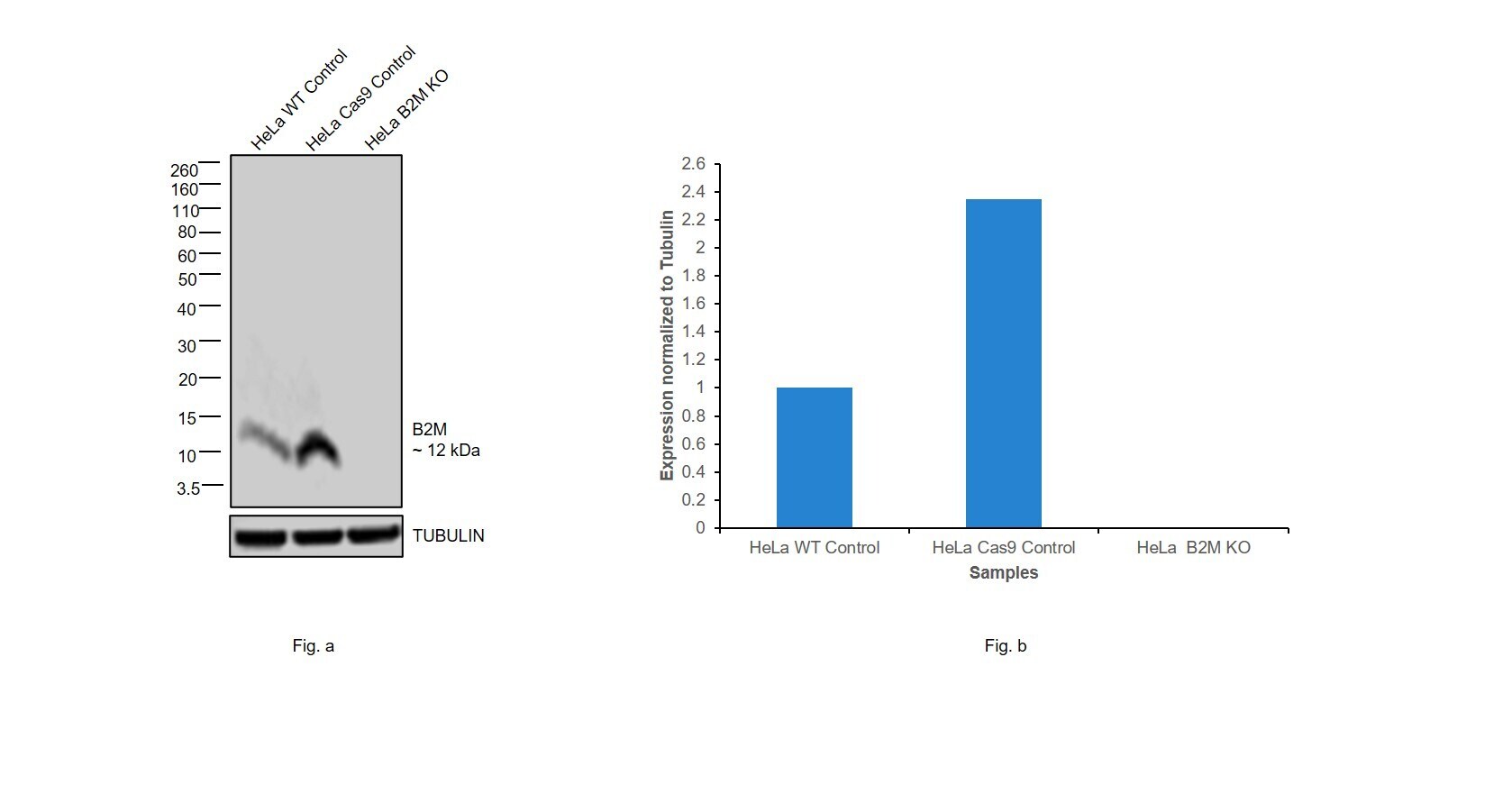
- Knockout of B2M was achieved by CRISPR-Cas9 genome editing using LentiArray™ Lentiviral sgRNA (Product # A32042, Assay ID CRISPR983707_LV) and LentiArray Cas9 Lentivirus (Product # A32064). Western blot analysis of B2M was performed by loading 50 µg of HeLa Wild Type (Lane 1), HeLa Cas9 (Lane 2) andHeLa B2M KO (Lane 3) membrane enriched extracts. The samples were electrophoresed using Novex™ 16%, Tricine, 1.0 mm, Mini Protein Gel (Product # EC66952BOX). Resolved proteins were then transferred onto a nitrocellulose membrane (Product # IB23001) by iBlot® 2 Dry Blotting System (Product # IB21001). The blot was probed with Anti-beta-2 Microglobulin Monoclonal Antibody (B2M-01) (Product # MA1-19141, 5 µg/mL dilution) and Goat anti-Mouse IgG (H+L) Superclonal™ Recombinant Secondary Antibody, HRP (Product # A28177, 1:5,000 dilution) using the iBright FL 1000 (Product # A32752). Chemiluminescent detection was performed using SuperSignal™ West Atto Ultimate Sensitivity Substrate (Product # A38556). Loss of signal upon CRISPR mediated knockout (KO) using the LentiArray™ CRISPR product line confirms that antibody is specific to B2M.
- Submitted by
- Invitrogen Antibodies (provider)
- Main image
- Experimental details
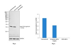
- Knockdown of beta-2 Microglobulin was achieved by transfecting HeLa with beta-2 Microglobulin specific siRNAs (Silencer® select Product # s1854, s1853). Western blot analysis (Fig. a) was performed using Whole cell extracts from the beta-2 Microglobulin knockdown cells (lane 3), non-targeting scrambled siRNA transfected cells (lane 2) and untransfected cells (lane 1). The blot was probed with beta-2 Microglobulin Monoclonal Antibody (B2M-01) (Product # MA1-19141, 3 microgram/ml) and Goat anti-Mouse IgG (H+L) Superclonal™ Recombinant Secondary Antibody, HRP (Product # A28177, 1:4000 dilution). Densitometric analysis of this western blot is shown in histogram (Fig. b). Decrease in signal upon siRNA mediated knock down confirms that antibody is specific to beta-2 Microglobulin.

- Submitted by
- Invitrogen Antibodies (provider)
- Main image

- Experimental details
- Western blot was performed using Anti-beta-2 Microglobulin Monoclonal Antibody (B2M-01) (Product # MA1-19141) and a 12 kDa band corresponding to beta-2 Microglobulin was observed across all cell lines tested except for SH-SY5Y and NTERA2 cl.D1, which are reported to express low levels of beta-2 Microglobulin. Whole cell extracts (30 µg lysate) of HeLa (Lane 1), THP-1 (Lane 2), U-937 (Lane 3), SH-SY5Y (Lane 4) and NTERA-2 cl.D1 (Lane 5) were electrophoresed using Novex™ 16% Tricine Protein Gel (Product # EC6695BOX). Resolved proteins were then transferred onto a Nitrocellulose membrane (Product # IB23001) by iBlot® 2 Dry Blotting System (Product # IB21001). The blot was probed with the primary antibody (3 µg/mL) and detected by chemiluminescence with Goat anti-Mouse IgG (H+L) Superclonal™ Recombinant Secondary Antibody, HRP (Product # A28177,1:4000 dilution) using the iBright FL1000 (Product # A32752). Chemiluminescent detection was performed using SuperSignal™ West Dura Extended Duration Substrate (Product # 34076).
