 Explore
 Validate
 Learn
Learn Western blot
 Immunocytochemistry
Antibody data
- Antibody Data
- Antigen structure
- References [0]
- Comments [0]
- Validations
- Western blot [2]
- Flow cytometry [1]
Submit
Validation data
Reference
Comment
Report error
- Product number
- MAB8480 - Provider product page

- Provider
- Novus Biologicals
- Product name
- Rabbit Monoclonal EPS15 Antibody
- Antibody type
- Monoclonal
- Description
- Protein A or G purified from cell culture supernatant. Detects human Eps15 in direct ELISAs and Western blots.
- Reactivity
- Human
- Host
- Rabbit
- Conjugate
- Unconjugated
- Isotype
- IgG
- Vial size
- 100 ug
- Storage
- Use a manual defrost freezer and avoid repeated freeze-thaw cycles. 12 months from date of receipt, -20 to -70 degreesC as supplied. 1 month, 2 to 8 degreesC under sterile conditions after reconstitution. 6 months, -20 to -70 degreesC under sterile conditions after reconstitution.
No comments: Submit comment
Supportive validation
- Submitted by
- Novus Biologicals (provider)
- Main image

- Experimental details
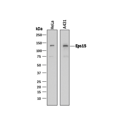
- Detection of Human Eps15 by Western Blot. Western blot shows lysates of HeLa human cervical epithelial carcinoma cell line and A431 human epithelial carcinoma cell line. PVDF membrane was probed with 0.1 µg/mL of Rabbit Anti-Human Eps15 Monoclonal Antibody (Catalog # MAB8480) followed by HRP-conjugated Anti-Rabbit IgG Secondary Antibody (Catalog # HAF008). A specific band was detected for Eps15 at approximately 140 kDa (as indicated). This experiment was conducted under reducing conditions and using Immunoblot Buffer Group 1.
- Submitted by
- Novus Biologicals (provider)
- Main image

- Experimental details
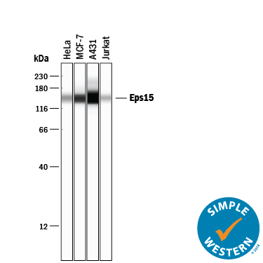
- Detection of Human Eps15 by Simple WesternTM. Simple Western lane view shows lysates of HeLa human cervical epithelial carcinoma cell line, MCF-7 human breast cancer cell line, A431 human epithelial carcinoma cell line, and Jurkat human acute T cell leukemia cell line, loaded at 0.2 mg/mL. A specific band was detected for Eps15 at approximately 150 kDa (as indicated) using 1 µg/mL of Rabbit Anti-Human Eps15 Monoclonal Antibody (Catalog # MAB8480). This experiment was conducted under reducing conditions and using the 12-230 kDa separation system.
Supportive validation
- Submitted by
- Novus Biologicals (provider)
- Main image
- Experimental details
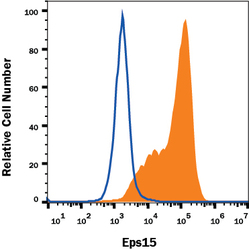
- Detection of Eps15 in U-118-MG Human Cell Line by Flow Cytometry. U-118-MG human glioblastoma/astrocytoma cell line was stained with Rabbit Anti-Human Eps15 Monoclonal Antibody (Catalog # MAB8480, filled histogram) or isotype control antibody (Catalog # AB-105-C, open histogram), followed by Allophycocyanin-conjugated Anti-Rabbit IgG Secondary Antibody (Catalog # F0111). To facilitate intracellular staining, cells were fixed and permeabilized with FlowX FoxP3 Fixation & Permeabilization Buffer Kit (Catalog # FC012). View our protocol for Staining Intracellular Molecules.
