Explore
 Validate
 Learn
Learn Western blot
 ELISA
ELISAAntibody data
- Antibody Data
- Antigen structure
- References [37]
- Comments [0]
- Validations
- Western blot [1]
- Immunocytochemistry [1]
- Immunohistochemistry [2]
Submit
Validation data
Reference
Comment
Report error
- Product number
- 16036-1-AP - Provider product page

- Provider
- Proteintech Group
- Proper citation
- Proteintech Cat#16036-1-AP, RRID:AB_2227602
- Product name
- Amphiregulin antibody
- Antibody type
- Polyclonal
- Description
- KD/KO validated Amphiregulin antibody (Cat. #16036-1-AP) is a rabbit polyclonal antibody that shows reactivity with human and has been validated for the following applications: FC, IF, IHC, IP, WB,ELISA.
- Reactivity
- Human
- Host
- Rabbit
- Conjugate
- Unconjugated
- Isotype
- IgG
- Vial size
- 20ul, 150ul
Submitted references O-GlcNAcylation of YAP1 promotes lung transplant ischemia-reperfusion injury via binding to HIF1α transcription factor and activating autophagy and mitophagy.
Amphiregulin and Epiregulin Confer Radioresistance in Esophageal Squamous Cell Carcinoma Through Oxidative Phosphorylation.
EGCG-releasing nanofibrous scaffold enhances wound healing via γδ T cells modulation.
Targeting Egfr-Mediated Cell Proliferation and Lipid Metabolism Separation Effectively Accelerate Liver Regeneration.
Brigatinib can inhibit proliferation and induce apoptosis of human immortalized keratinocyte cells.
Amphiregulin Mediates Epithelial Cell-Eosinophil Interactions and Amplifies Inflammation in Chronic Rhinosinusitis With Nasal Polyps.
17β-Estradiol Promotes Tumorigenicity Through an Autocrine AREG/EGFR Loop in ER-α-Positive Breast Cancer Cells.
Pannexin1 via P2rx7/amphiregulin contributes to cardiac fibrosis post myocardial infarction.
BMAL1 ameliorates type 2 diabetes-induced cognitive impairment via AREG upregulation and PI3K/Akt/GSK-3β pathway activation.
Multi-omic analysis of gallbladder cancer identifies distinct tumor microenvironments associated with disease progression.
Integrated multi-omics profiling highlights the diet-gut-brain axis in low-calorie diets promoted novelty-seeking behavior.
Silencing AREG Enhances Sensitivity to Irradiation by Suppressing the PI3K/AKT Signaling Pathway in Colorectal Cancer Cells.
Enhanced amphiregulin exposure promotes modulation of the high grade serous ovarian cancer tumor immune microenvironment.
Hyodeoxycholic acid inhibits colorectal cancer proliferation through the FXR/EREG/EGFR axis.
Liver X Receptors Enhance Epithelial to Mesenchymal Transition in Metastatic Prostate Cancer Cells.
TLR9 activation in large wound induces tissue repair and hair follicle regeneration via γδT cells.
Jiawei Danxuan Koukang Alleviates Arecoline Induced Oral Mucosal Lesions: Network Pharmacology and the Combined Ultra-High Performance Liquid Chromatography (UPLC) and Mass Spectrometry (MS).
Amphiregulin blockade decreases the levodopa-induced dyskinesia in a 6-hydroxydopamine Parkinson's disease mouse model.
Dapagliflozin delays renal fibrosis in diabetic kidney disease by inhibiting YAP/TAZ activation.
Quercetin inhibits the amphiregulin/EGFR signaling-mediated renal tubular epithelial-mesenchymal transition and renal fibrosis in obstructive nephropathy.
TPM2 attenuates progression of prostate cancer by blocking PDLIM7-mediated nuclear translocation of YAP1.
Schwann cell-derived amphiregulin enhances nerve regeneration via supporting the proliferation and migration of Schwann cells and the elongation of axons.
miR-33a-3p regulates METTL3-mediated AREG stability and alters EMT to inhibit pancreatic cancer invasion and metastasis.
Adaptive transcriptomic and immune infiltrate responses in the tumor immune microenvironment following neoadjuvant chemotherapy in high grade serous ovarian cancer reveal novel prognostic associations and activation of pro-tumorigenic pathways.
Establishment of a patient-derived mucoepidermoid carcinoma cell line with the CRTC1-MAML2 fusion gene.
Luteinizing hormone stimulates the expression of amphiregulin in human theca cells.
AXL cooperates with EGFR to mediate neutrophil elastase-induced migration of prostate cancer cells.
Extracellular Matrix-Bound FGF2 Mediates Estrogen Receptor Signaling and Therapeutic Response in Breast Cancer.
IFI16 promotes cervical cancer progression by upregulating PD-L1 in immunomicroenvironment through STING-TBK1-NF-kB pathway.
CBX2 Regulates Proliferation and Apoptosis via the Phosphorylation of YAP in Hepatocellular Carcinoma.
CD317 Activates EGFR by Regulating Its Association with Lipid Rafts.
AhR controls redox homeostasis and shapes the tumor microenvironment in BRCA1-associated breast cancer.
Targeting amphiregulin (AREG) derived from senescent stromal cells diminishes cancer resistance and averts programmed cell death 1 ligand (PD-L1)-mediated immunosuppression.
Loss of KIBRA function activates EGFR signaling by inducing AREG.
Membrane-Tethered Intracellular Domain of Amphiregulin Promotes Keratinocyte Proliferation.
The EGF receptor ligand amphiregulin controls cell division via FoxM1.
TAZ induces growth factor-independent proliferation through activation of EGFR ligand amphiregulin.
Dai S, Wan X, Xia L, Xu L, Xie C, Wang G, Tang J
Cell death & disease 2026 Mar 15;17(1)
Cell death & disease 2026 Mar 15;17(1)
Amphiregulin and Epiregulin Confer Radioresistance in Esophageal Squamous Cell Carcinoma Through Oxidative Phosphorylation.
Lin Z, Yao M, Xu X, Zhang D, Xu L, Rong L, Wang X, Duan Y, Chen C, Gu J, Zhang Y, Liu Q, Ye Q, Cui G, Hao Y, Ma X
Advanced science (Weinheim, Baden-Wurttemberg, Germany) 2026 Jan;13(6):e07524
Advanced science (Weinheim, Baden-Wurttemberg, Germany) 2026 Jan;13(6):e07524
EGCG-releasing nanofibrous scaffold enhances wound healing via γδ T cells modulation.
An T, Xu Z, Yang Y, Li X, Chen S, Yang X, Man Y, Hu C, Qu Y
Biomaterials 2026 Feb;325:123543
Biomaterials 2026 Feb;325:123543
Targeting Egfr-Mediated Cell Proliferation and Lipid Metabolism Separation Effectively Accelerate Liver Regeneration.
Hu Y, Song S, Wang R, An N, Diao J, Chen Y, Liu J, Lv G
Cell proliferation 2026 Apr 22;:e70214
Cell proliferation 2026 Apr 22;:e70214
Brigatinib can inhibit proliferation and induce apoptosis of human immortalized keratinocyte cells.
Yang Q, Zhao D, Ju L, Cao P, Wei J, Liu Z
Frontiers in pharmacology 2025;16:1524277
Frontiers in pharmacology 2025;16:1524277
Amphiregulin Mediates Epithelial Cell-Eosinophil Interactions and Amplifies Inflammation in Chronic Rhinosinusitis With Nasal Polyps.
Zhao L, Zhang S, Zhang Y, Liu Y, Guo Y, Li Y, Wang Q, Wang Z, Qu Z, Zhang N, Bachert C, Wang C, Zhang L, Lan F
Allergy 2025 May;80(5):1335-1347
Allergy 2025 May;80(5):1335-1347
17β-Estradiol Promotes Tumorigenicity Through an Autocrine AREG/EGFR Loop in ER-α-Positive Breast Cancer Cells.
Yoon SY, Jeong Y, Ryu JM, Lee SK, Chae BJ, Yu J, Kim SW, Nam SJ, Kim S, Lee JE
Cells 2025 May 12;14(10)
Cells 2025 May 12;14(10)
Pannexin1 via P2rx7/amphiregulin contributes to cardiac fibrosis post myocardial infarction.
Deng N, You L, Guo H, Wei Y, Xu F, Chen D, Luo S, Huang S, Zuo S, Li W, Si X
Journal of molecular histology 2025 Jul 15;56(4):230
Journal of molecular histology 2025 Jul 15;56(4):230
BMAL1 ameliorates type 2 diabetes-induced cognitive impairment via AREG upregulation and PI3K/Akt/GSK-3β pathway activation.
Xu J, Li C, Fan R, Yin J, Xie L, Peng X, Tao J, Xu W, Zhang S, Shi X, Dong K, Yu X, Chen X, Yang Y
Cell communication and signaling : CCS 2025 Jan 6;23(1):7
Cell communication and signaling : CCS 2025 Jan 6;23(1):7
Multi-omic analysis of gallbladder cancer identifies distinct tumor microenvironments associated with disease progression.
Zhou T, Wu Y, Li S, Qiu X, Liu E, Xie Z, Shi X, Zhang Y, Ma G, Guo W, Wang X, Wang K, Yao X, Hu J, Shen S, Yang S, Jiang X, Fu J, Wang H, Gu J, Chen L
Nature genetics 2025 Aug;57(8):1935-1949
Nature genetics 2025 Aug;57(8):1935-1949
Integrated multi-omics profiling highlights the diet-gut-brain axis in low-calorie diets promoted novelty-seeking behavior.
Wang S, Su LY, Chen J, Tian Y, Zhou H
Current research in food science 2024;9:100897
Current research in food science 2024;9:100897
Silencing AREG Enhances Sensitivity to Irradiation by Suppressing the PI3K/AKT Signaling Pathway in Colorectal Cancer Cells.
Zhang W, Zhang W, Tang C, Hu Y, Yi K, Xu X, Chen Z
Biologics : targets & therapy 2024;18:273-284
Biologics : targets & therapy 2024;18:273-284
Enhanced amphiregulin exposure promotes modulation of the high grade serous ovarian cancer tumor immune microenvironment.
Ebott J, McAdams J, Kim C, Jansen C, Woodman M, De La Cruz P, Schrol C, Ribeiro J, James N
Frontiers in pharmacology 2024;15:1375421
Frontiers in pharmacology 2024;15:1375421
Hyodeoxycholic acid inhibits colorectal cancer proliferation through the FXR/EREG/EGFR axis.
Pang Q, Huang S, Li X, Cao J
Frontiers in cell and developmental biology 2024;12:1480998
Frontiers in cell and developmental biology 2024;12:1480998
Liver X Receptors Enhance Epithelial to Mesenchymal Transition in Metastatic Prostate Cancer Cells.
Bouchareb E, Dallel S, De Haze A, Damon-Soubeyrand C, Renaud Y, Baabdaty E, Vialat M, Fabre J, Pouchin P, De Joussineau C, Degoul F, Sanmukh S, Gendronneau J, Sanchez P, Gonthier-Gueret C, Trousson A, Morel L, Lobaccaro JM, Kocer A, Baron S
Cancers 2024 Aug 6;16(16)
Cancers 2024 Aug 6;16(16)
TLR9 activation in large wound induces tissue repair and hair follicle regeneration via γδT cells.
Li X, An T, Yang Y, Xu Z, Chen S, Yi Z, Deng C, Zhou F, Man Y, Hu C
Cell death & disease 2024 Aug 17;15(8):598
Cell death & disease 2024 Aug 17;15(8):598
Jiawei Danxuan Koukang Alleviates Arecoline Induced Oral Mucosal Lesions: Network Pharmacology and the Combined Ultra-High Performance Liquid Chromatography (UPLC) and Mass Spectrometry (MS).
Zhou L, Tan J, Dai Y, Zhu K, Xiao Y, Wu D, Wang Z, Tan Y, Qin Y
Drug design, development and therapy 2023;17:3085-3101
Drug design, development and therapy 2023;17:3085-3101
Amphiregulin blockade decreases the levodopa-induced dyskinesia in a 6-hydroxydopamine Parkinson's disease mouse model.
Kambey PA, Liu WY, Wu J, Tang C, Buberwa W, Saro A, Nyalali AMK, Gao D
CNS neuroscience & therapeutics 2023 Oct;29(10):2925-2939
CNS neuroscience & therapeutics 2023 Oct;29(10):2925-2939
Dapagliflozin delays renal fibrosis in diabetic kidney disease by inhibiting YAP/TAZ activation.
Feng L, Chen Y, Li N, Yang X, Zhou L, Li H, Wang T, Xie M, Liu H
Life sciences 2023 Jun 1;322:121671
Life sciences 2023 Jun 1;322:121671
Quercetin inhibits the amphiregulin/EGFR signaling-mediated renal tubular epithelial-mesenchymal transition and renal fibrosis in obstructive nephropathy.
Wang Q, Wang F, Li X, Ma Z, Jiang D
Phytotherapy research : PTR 2023 Jan;37(1):111-123
Phytotherapy research : PTR 2023 Jan;37(1):111-123
TPM2 attenuates progression of prostate cancer by blocking PDLIM7-mediated nuclear translocation of YAP1.
Wu Z, Ge L, Ma L, Lu M, Song Y, Deng S, Duan P, Du T, Wu Y, Zhang Z, Zhang S
Cell & bioscience 2023 Feb 23;13(1):39
Cell & bioscience 2023 Feb 23;13(1):39
Schwann cell-derived amphiregulin enhances nerve regeneration via supporting the proliferation and migration of Schwann cells and the elongation of axons.
Chen S, Chen Q, Zhang X, Shen Y, Shi X, Dai X, Yi S
Journal of neurochemistry 2023 Aug;166(4):678-691
Journal of neurochemistry 2023 Aug;166(4):678-691
miR-33a-3p regulates METTL3-mediated AREG stability and alters EMT to inhibit pancreatic cancer invasion and metastasis.
Su X, Lai T, Tao Y, Zhang Y, Zhao C, Zhou J, Chen E, Zhu M, Zhang S, Wang B, Mao Y, Hu H
Scientific reports 2023 Aug 21;13(1):13587
Scientific reports 2023 Aug 21;13(1):13587
Adaptive transcriptomic and immune infiltrate responses in the tumor immune microenvironment following neoadjuvant chemotherapy in high grade serous ovarian cancer reveal novel prognostic associations and activation of pro-tumorigenic pathways.
James NE, Woodman M, De La Cruz P, Eurich K, Ozsoy MA, Schorl C, Hanley LC, Ribeiro JR
Frontiers in immunology 2022;13:965331
Frontiers in immunology 2022;13:965331
Establishment of a patient-derived mucoepidermoid carcinoma cell line with the CRTC1-MAML2 fusion gene.
Noguchi K, Kanda S, Yoshida K, Funaoka Y, Yamanegi K, Yoshikawa K, Takaoka K, Kishimoto H, Nakano Y
Molecular and clinical oncology 2022 Mar;16(3):75
Molecular and clinical oncology 2022 Mar;16(3):75
Luteinizing hormone stimulates the expression of amphiregulin in human theca cells.
Liu Y, Zhong Y, Shen X, Guo X, Wu R, Yang T, Chen M
Journal of ovarian research 2022 Dec 7;15(1):129
Journal of ovarian research 2022 Dec 7;15(1):129
AXL cooperates with EGFR to mediate neutrophil elastase-induced migration of prostate cancer cells.
Xiao Z, Hammes SR
iScience 2021 Nov 19;24(11):103270
iScience 2021 Nov 19;24(11):103270
Extracellular Matrix-Bound FGF2 Mediates Estrogen Receptor Signaling and Therapeutic Response in Breast Cancer.
DiGiacomo JW, Godet I, Trautmann-Rodriguez M, Gilkes DM
Molecular cancer research : MCR 2021 Jan;19(1):136-149
Molecular cancer research : MCR 2021 Jan;19(1):136-149
IFI16 promotes cervical cancer progression by upregulating PD-L1 in immunomicroenvironment through STING-TBK1-NF-kB pathway.
Cai H, Yan L, Liu N, Xu M, Cai H
Biomedicine & pharmacotherapy = Biomedecine & pharmacotherapie 2020 Mar;123:109790
Biomedicine & pharmacotherapy = Biomedecine & pharmacotherapie 2020 Mar;123:109790
CBX2 Regulates Proliferation and Apoptosis via the Phosphorylation of YAP in Hepatocellular Carcinoma.
Mao J, Tian Y, Wang C, Jiang K, Li R, Yao Y, Zhang R, Sun D, Liang R, Gao Z, Wang Q, Wang L
Journal of Cancer 2019;10(12):2706-2719
Journal of Cancer 2019;10(12):2706-2719
CD317 Activates EGFR by Regulating Its Association with Lipid Rafts.
Zhang G, Li X, Chen Q, Li J, Ruan Q, Chen YH, Yang X, Wan X
Cancer research 2019 May 1;79(9):2220-2231
Cancer research 2019 May 1;79(9):2220-2231
AhR controls redox homeostasis and shapes the tumor microenvironment in BRCA1-associated breast cancer.
Kubli SP, Bassi C, Roux C, Wakeham A, Göbl C, Zhou W, Jafari SM, Snow B, Jones L, Palomero L, Thu KL, Cassetta L, Soong D, Berger T, Ramachandran P, Baniasadi SP, Duncan G, Lindzen M, Yarden Y, Herranz C, Lazaro C, Chu MF, Haight J, Tinto P, Silvester J, Cescon DW, Petit A, Pettersson S, Pollard JW, Mak TW, Pujana MA, Cappello P, Gorrini C
Proceedings of the National Academy of Sciences of the United States of America 2019 Feb 26;116(9):3604-3613
Proceedings of the National Academy of Sciences of the United States of America 2019 Feb 26;116(9):3604-3613
Targeting amphiregulin (AREG) derived from senescent stromal cells diminishes cancer resistance and averts programmed cell death 1 ligand (PD-L1)-mediated immunosuppression.
Xu Q, Long Q, Zhu D, Fu D, Zhang B, Han L, Qian M, Guo J, Xu J, Cao L, Chin YE, Coppé JP, Lam EW, Campisi J, Sun Y
Aging cell 2019 Dec;18(6):e13027
Aging cell 2019 Dec;18(6):e13027
Loss of KIBRA function activates EGFR signaling by inducing AREG.
Mussell AL, Denson KE, Shen H, Chen Y, Yang N, Frangou C, Zhang J
Oncotarget 2018 Jul 6;9(52):29975-29984
Oncotarget 2018 Jul 6;9(52):29975-29984
Membrane-Tethered Intracellular Domain of Amphiregulin Promotes Keratinocyte Proliferation.
Stoll SW, Stuart PE, Lambert S, Gandarillas A, Rittié L, Johnston A, Elder JT
The Journal of investigative dermatology 2016 Feb;136(2):444-452
The Journal of investigative dermatology 2016 Feb;136(2):444-452
The EGF receptor ligand amphiregulin controls cell division via FoxM1.
Stoll SW, Stuart PE, Swindell WR, Tsoi LC, Li B, Gandarillas A, Lambert S, Johnston A, Nair RP, Elder JT
Oncogene 2016 Apr 21;35(16):2075-86
Oncogene 2016 Apr 21;35(16):2075-86
TAZ induces growth factor-independent proliferation through activation of EGFR ligand amphiregulin.
Yang N, Morrison CD, Liu P, Miecznikowski J, Bshara W, Han S, Zhu Q, Omilian AR, Li X, Zhang J
Cell cycle (Georgetown, Tex.) 2012 Aug 1;11(15):2922-30
Cell cycle (Georgetown, Tex.) 2012 Aug 1;11(15):2922-30
No comments: Submit comment
Supportive validation
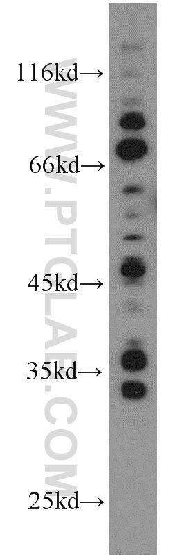
- Submitted by
- Proteintech Group (provider)
- Main image

- Experimental details
- MCF7 cells were subjected to SDS PAGE followed by western blot with 16036-1-AP(AREG antibody) at dilution of 1:2000
- Sample type
- cell line
Supportive validation
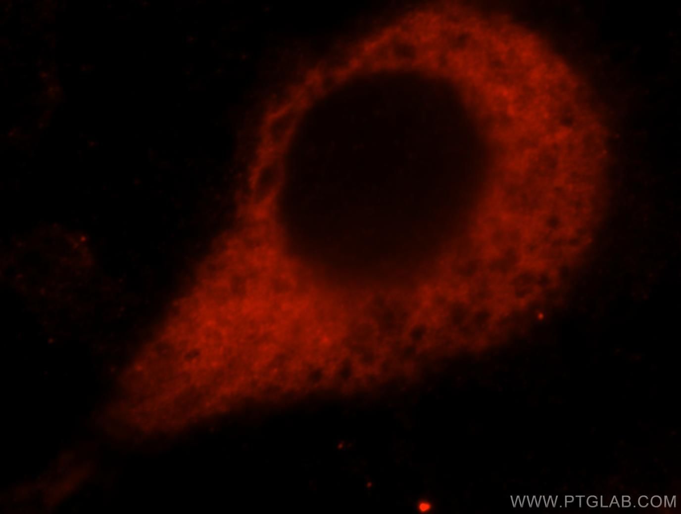
- Submitted by
- Proteintech Group (provider)
- Main image

- Experimental details
- Immunofluorescent analysis of Hela cells, using AREG antibody 16036-1-AP at 1:25 dilution and Rhodamine-labeled goat anti-rabbit IgG (red).
- Sample type
- cell line
Supportive validation
- Submitted by
- Proteintech Group (provider)
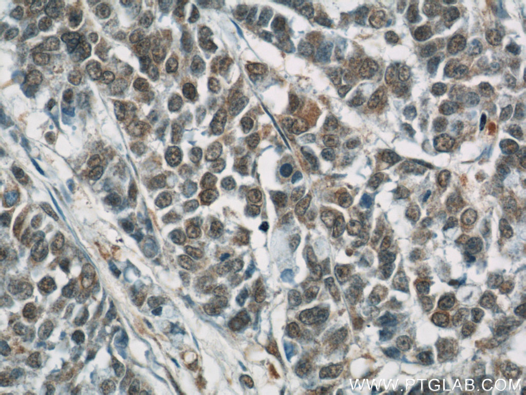
- Main image
- Experimental details
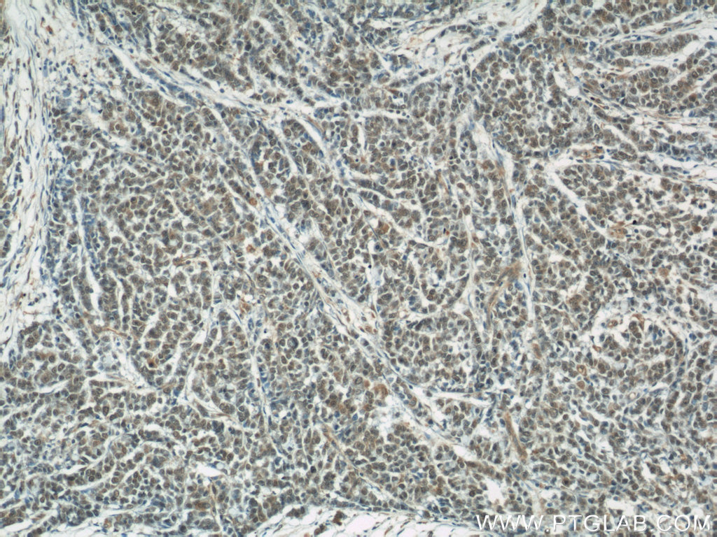
- Immunohistochemical of paraffin-embedded human colon cancer using 16036-1-AP(AREG antibody) at dilution of 1:50 (under 10x lens)
- Sample type
- tissue
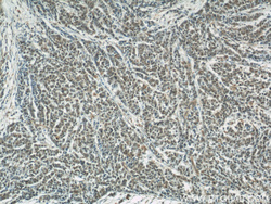
- Submitted by
- Proteintech Group (provider)
- Main image
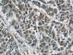
- Experimental details
- Immunohistochemical of paraffin-embedded human colon cancer using 16036-1-AP(AREG antibody) at dilution of 1:50 (under 40x lens)
- Sample type
- tissue