Explore
 Validate
 Learn
Learn Western blot
Antibody data
- Antibody Data
- Antigen structure
- References [1]
- Comments [0]
- Validations
- Western blot [1]
- Flow cytometry [2]
- Other assay [1]
Submit
Validation data
Reference
Comment
Report error
- Product number
- PA5-26137 - Provider product page

- Provider
- Invitrogen Antibodies
- Product name
- CLRN3 Polyclonal Antibody
- Antibody type
- Polyclonal
- Antigen
- Synthetic peptide
- Reactivity
- Human
- Host
- Rabbit
- Isotype
- IgG
- Vial size
- 400 μL
- Concentration
- 0.5 mg/mL
- Storage
- Store at 4°C short term. For long term storage, store at -20°C, avoiding freeze/thaw cycles.
Submitted references Mapping the Cell-Surface N-Glycoproteome of Human Hepatocytes Reveals Markers for Selecting a Homogeneous Population of iPSC-Derived Hepatocytes.
Mallanna SK, Cayo MA, Twaroski K, Gundry RL, Duncan SA
Stem cell reports 2016 Sep 13;7(3):543-556
Stem cell reports 2016 Sep 13;7(3):543-556
No comments: Submit comment
Supportive validation
- Submitted by
- Invitrogen Antibodies (provider)
- Main image

- Experimental details
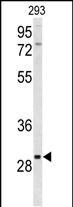
- Western blot analysis of CLRN3 in 293 cell line lysates. Samples were incubated with CLRN3 polyclonal antibody (Product # PA5-26137). Lysates: 35 µg/lane. CLRN3 (arrow).
Supportive validation
- Submitted by
- Invitrogen Antibodies (provider)
- Main image
- Experimental details
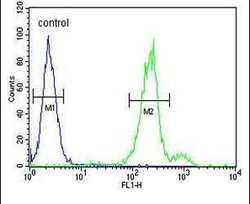
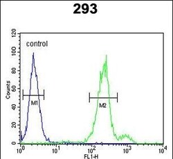
- Flow cytometry analysis of 293 cells using a CLRN3 polyclonal antibody (Product # PA5-26137) (right) compared to a negative control cell (left) at a dilution of 1:10-50, followed by a FITC-conjugated goat anti-rabbit antibody
- Submitted by
- Invitrogen Antibodies (provider)
- Main image
- Experimental details
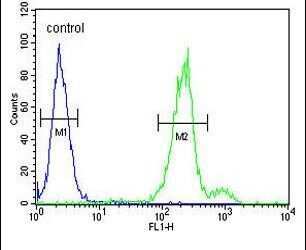
- Flow cytometry of CLRN3 in 293 cells (right histogram). Samples were incubated with CLRN3 polyclonal antibody (Product # PA5-26137) followed by FITC-conjugated goat-anti-rabbit secondary antibody. Negative control cell (left histogram).

Supportive validation
- Submitted by
- Invitrogen Antibodies (provider)
- Main image
- Experimental details
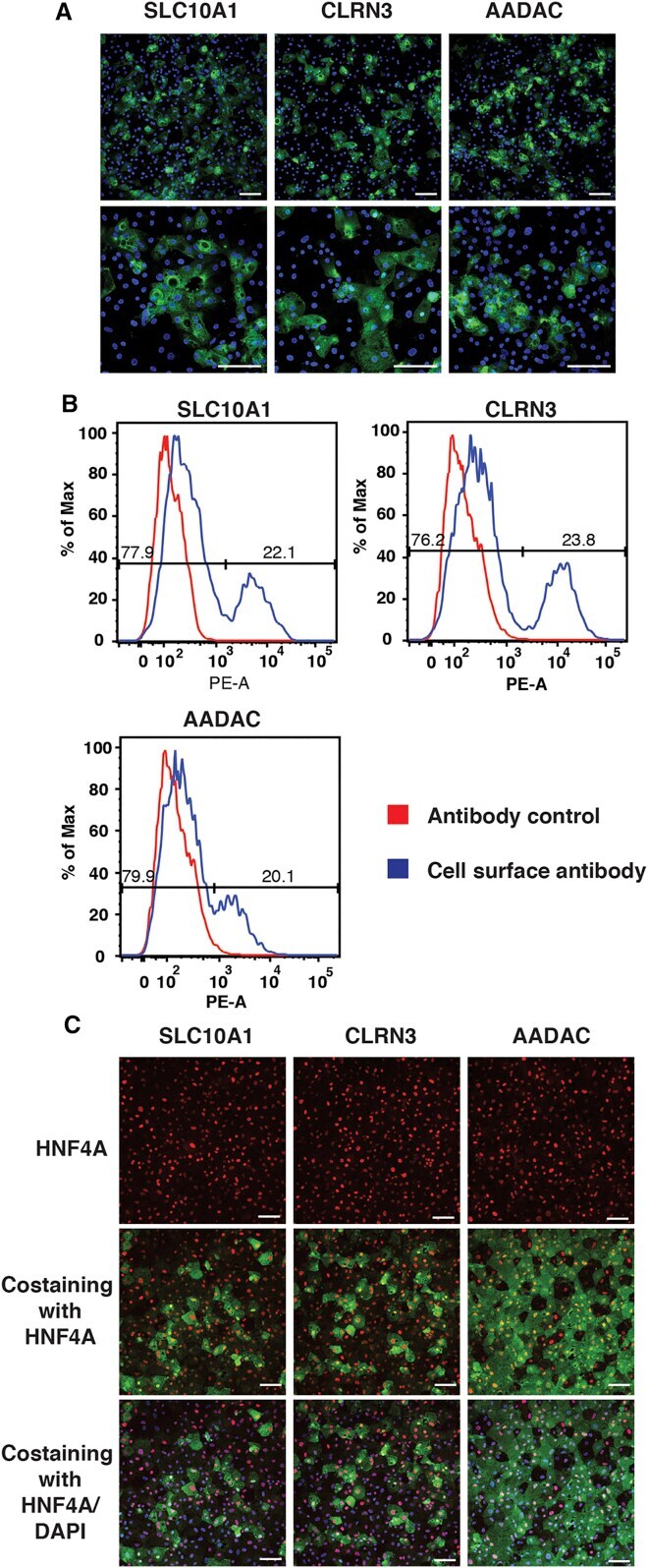
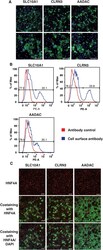
- Figure 4 A Subpopulation of iPSC-Derived Hepatocyte-like Cells Express SLC10A1, CLRN3, and AADAC (A) Confocal micrographs showing the results of immunocytochemistry to detect expression of cell-surface proteins SLC10A1, CLRN3, and AADAC in iPSC-derived hepatocyte-like cells (green). Nuclei are identified by DAPI staining (blue) (see also Figures S3 B and S3C). Scale bars, 100 mum. (B) FACS histogram plots of iPSC-derived hepatocyte-like cells stained with primary antibodies against proteins SLC10A1, CLRN3, and AADAC and corresponding phycoerythrin (PE) conjugated secondary antibody are shown. Cells stained with PE conjugated secondary antibody only are used as control. (C) Confocal micrographs showing the results of immunocytochemistry to detect expression of HNF4A (red) and cell-surface proteins SLC10A1, CLRN3, and AADAC (green) in iPSC-derived hepatocyte-like cells (see also Figure S4 ). Nuclei are identified by DAPI staining (blue). Scale bars, 100 mum.